A DEXA scan is a quick, painless test that measures how much mineral is present in certain bones. It is most commonly used to investigate osteoporosis, estimate the likelihood of future fractures and help doctors decide whether bone-strengthening treatment may be beneficial.
The test is also called a DXA scan, bone-density scan or bone densitometry scan. DEXA and DXA refer to the same examination: dual-energy X-ray absorptiometry.
Most scans measure the lower spine and hips because fractures in these areas can have a major effect on health, mobility and independence. The result is usually reported as a T-score and sometimes a Z-score, but these numbers are only part of the assessment. A person can have moderately reduced bone density and still be at high fracture risk because of age, previous fractures, steroid treatment or another medical condition.
A DEXA scan is not usually an emergency test. However, seek prompt medical advice if you develop sudden severe back pain, lose height noticeably, become increasingly stooped or break a bone after a minor fall. These may indicate a fragility fracture and should not be ignored while waiting for a routine bone-density appointment.
What is a DEXA scan?
A DEXA scan uses two very low-energy X-ray beams to estimate the mineral content of bone. Different tissues absorb the beams by different amounts, allowing the scanner’s software to calculate bone mineral density.
Bone mineral density is commonly abbreviated to BMD. The measurement is usually expressed in grams of mineral per square centimetre, although patients are more often shown the resulting T-score or Z-score than the raw density value.
In adults, the main areas scanned are usually:
- the lumbar spine in the lower back;
- the total hip;
- the femoral neck, which is the narrower upper part of the thigh bone near the hip joint.
Sometimes the forearm is scanned instead. This may be useful when the hip or spine cannot be measured accurately because of joint replacements, metalwork, severe arthritis, spinal surgery or another technical reason.
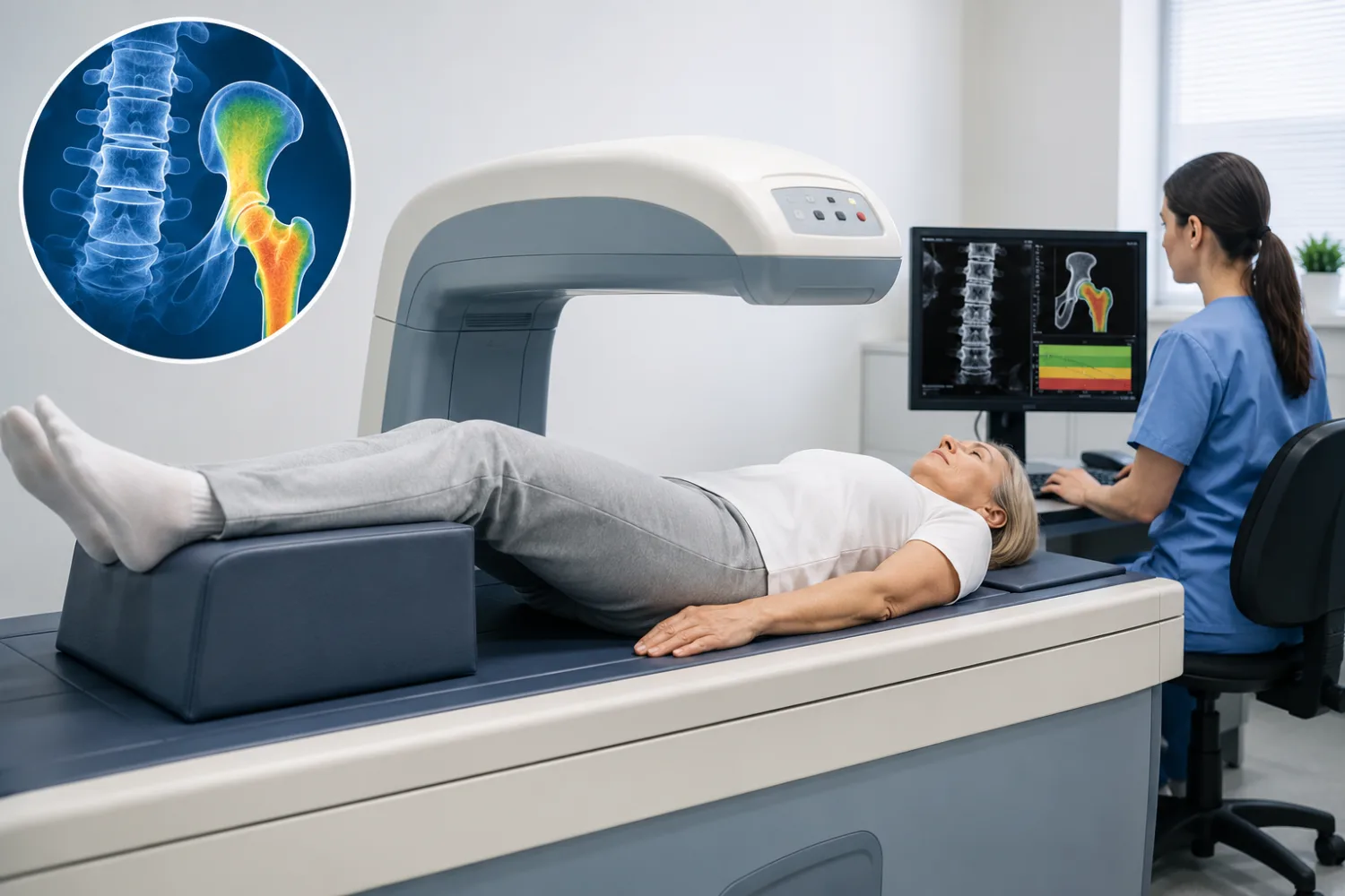
A DEXA scanner does not look like an enclosed MRI machine. It consists of a flat examination table with a scanning arm that moves over the body. Nothing normally touches or presses on you during the scan.
Is it DEXA or DXA?
Both terms are widely used. DXA is the technically preferred abbreviation for dual-energy X-ray absorptiometry, while DEXA remains the term many patients and healthcare services recognise more easily.
They refer to the same bone-density test.
What does a bone-density scan actually show?
A DEXA scan shows whether the measured bones contain more or less mineral than expected when compared with reference populations.
In general, bones with lower mineral density are less resistant to fracture. However, density is not the same as total bone strength. Bone strength is also influenced by:
- the internal structure and quality of the bone;
- age;
- previous fractures;
- how quickly bone is being lost;
- falls risk;
- medical conditions;
- medicines;
- smoking and alcohol use;
- body weight and muscle strength.
This is why a DEXA result should not be read in isolation. Two people with the same T-score may have very different chances of breaking a bone.
Can a DEXA scan diagnose osteoporosis?
In postmenopausal women and men aged 50 or over, a sufficiently low T-score can support a diagnosis of osteoporosis.
Doctors can also diagnose and treat osteoporosis in some people because they have already experienced a typical fragility fracture, even when a scan result does not fall below the usual osteoporosis threshold.
A fragility fracture is a break caused by a force that would not normally be expected to fracture healthy bone, such as falling from standing height. Common sites include the wrist, hip, spine and upper arm.
Can it show a spinal fracture?
A standard bone-density scan is designed primarily to measure density, not to provide a detailed diagnostic image of every vertebra.
Some DEXA services can perform a separate low-radiation test called vertebral fracture assessment. This looks at the shape of the spine and may identify collapsed or compressed vertebrae.
A conventional X-ray, MRI or CT scan may still be needed when a new spinal fracture, nerve compression or another cause of back pain is suspected.
Can DEXA measure body fat?
A whole-body DEXA scan can estimate bone mass, fat mass and lean tissue. This type of body-composition scan is different from the standard hip-and-spine examination used to assess osteoporosis.
Whole-body DEXA is used in some specialist paediatric services, sports settings and research. It is not routinely required to assess an adult’s fracture risk.
Who may be offered a DEXA scan?
DEXA scans are not automatically offered to every adult at a particular birthday. In the UK, clinicians usually consider a person’s overall fracture risk before deciding whether bone-density measurement would add useful information.
You may be offered a scan if you have:
- broken a bone after a minor fall or relatively low level of force;
- risk factors for osteoporosis or fragility fractures;
- taken steroid tablets for a prolonged period;
- experienced early menopause;
- very low body weight;
- lost height or developed increasing spinal curvature;
- a condition that can weaken bones;
- a treatment that lowers sex-hormone levels;
- an unclear fracture-risk assessment where BMD would help guide treatment;
- started osteoporosis treatment that requires future monitoring.
Age and menopause
Bone density generally reaches its peak during early adulthood and then gradually decreases. Bone loss accelerates around and after menopause because oestrogen levels fall.
Older postmenopausal women are therefore at increased risk, but osteoporosis is not solely a women’s condition. Men also lose bone with age and can develop serious fragility fractures.
A younger woman may be assessed when menopause occurs unusually early, both ovaries have been removed, periods stop for a prolonged period or another medical condition affects hormone production.
Steroid medicines
Long-term use of oral corticosteroids such as prednisolone can reduce bone formation and increase fracture risk. Risk depends on the dose, duration, age and other individual factors.
People expected to take steroids for several months may need fracture-risk assessment, preventative treatment or a DEXA scan rather than waiting for symptoms to develop.
Do not stop steroid medicine suddenly. Abrupt withdrawal can be dangerous after prolonged use and should be managed by the prescribing clinician.
Conditions associated with lower bone density
Conditions that may increase the likelihood of osteoporosis include:
- rheumatoid arthritis and some other inflammatory diseases;
- coeliac disease and inflammatory bowel disease;
- overactive thyroid or parathyroid disorders;
- type 1 diabetes;
- chronic kidney or liver disease;
- malabsorption and some weight-loss surgery;
- eating disorders;
- low testosterone;
- multiple myeloma and some other cancers;
- long periods of immobility.
The presence of one condition does not automatically mean that osteoporosis is present. It means the person’s risk may deserve closer assessment.
Cancer treatments
Some breast-cancer and prostate-cancer treatments reduce oestrogen or testosterone levels and can accelerate bone loss. Oncology teams may arrange a baseline scan and subsequent monitoring depending on the treatment and other risk factors.
What happens during a DEXA scan?
A standard DEXA scan is quick, non-invasive and painless. The appointment may take around 10 to 30 minutes, while the actual scanning time can be shorter.
At the appointment, a radiographer or trained technician may:
- confirm your identity and reason for referral;
- ask about pregnancy possibility;
- review previous fractures, operations and implants;
- measure your height and weight;
- ask you to remove clothing or objects containing metal.
You will usually lie on your back on a flat padded table. The scanning arm moves slowly above the body while you remain still and breathe normally.
Scanning the spine
For the lower-spine measurement, your legs may be placed on a padded support. This helps flatten the natural curve of the lower back and improves positioning.
Scanning the hip
For the hip measurement, the leg may be gently turned inwards and held in position using a support. This allows consistent measurement of the femoral neck and total hip.
The position can feel unfamiliar, but it should not normally be painful. Tell the radiographer if arthritis, recent surgery or another condition makes positioning difficult.
Do you go inside a tunnel?
No. The scanner is open and the scanning arm passes above you. People who feel claustrophobic in MRI scanners usually tolerate DEXA without difficulty.
Will you need an injection?
No injection, contrast dye or sedative is normally needed for an ordinary bone-density scan.
Can you eat beforehand?
Most services do not require fasting. You can generally eat, drink and take your usual medicines unless your appointment letter says otherwise.
Local instructions vary, particularly around calcium supplements and recent contrast examinations, so follow the information provided by the scanning department.
How should you prepare for the scan?
DEXA usually requires very little preparation, but a few practical steps can reduce delays and improve accuracy.
It may help to:
- wear loose, comfortable clothing without metal zips, hooks or buckles;
- leave metal jewellery and removable accessories at home;
- bring details of previous fractures and osteoporosis medicines;
- tell the department about hip replacements or spinal metalwork;
- mention any recent barium study, nuclear-medicine test or contrast scan;
- tell staff if you are or could be pregnant.
Calcium supplements
Some scanning departments ask patients not to take calcium supplements on the morning of the scan because an undissolved tablet could potentially appear over the spine.
Other departments do not require this. Follow the instructions in your appointment letter rather than stopping supplements for several days without advice.
Previous scans
Bring previous reports if they were performed by another provider. Comparison is most reliable when repeat scans use the same machine, body sites and positioning method.
Small differences between machines can make minor changes difficult to interpret.
Pregnancy
DEXA uses a very low radiation dose, but it is usually postponed during pregnancy unless there is an exceptional clinical reason.
Tell the department before the scan if pregnancy is possible. This allows the team to decide whether the appointment should proceed, be delayed or be replaced by another form of assessment.
Understanding the T-score
The T-score compares your measured bone density with the average bone density of a healthy young adult of the same sex.
The result is expressed in standard deviations, showing how far your result sits above or below the young-adult average.
The commonly used categories are:
- above or equal to −1.0: bone density within the expected normal range;
- between −1.0 and −2.5: low bone density, often called osteopenia;
- −2.5 or lower: bone density in the osteoporosis range.
A result of −2.5 is lower than −2.0. As the number becomes more negative, measured bone density is lower.
Which T-score matters?
A report may include separate scores for:
- the lumbar spine;
- the total hip;
- the femoral neck;
- sometimes the forearm.
The lowest valid score is often clinically important, but the interpretation may be more complicated when arthritis, spinal degeneration, fractures or metalwork affect one site.
For example, age-related arthritis can make the lumbar spine appear artificially denser. A hip measurement may then provide a more reliable indication of bone density.
Does a T-score of −2.5 mean your bones will break?
No. A T-score does not predict with certainty whether an individual bone will fracture.
It indicates that density is in a range associated with a higher statistical risk. Some people with osteoporosis-range scores never fracture, while others break a bone with a score in the osteopenia range because they have additional risk factors.
Is osteopenia a disease?
Osteopenia means bone density is below the young-adult reference range but not low enough to meet the conventional DEXA definition of osteoporosis.
It does not automatically mean treatment is needed. Many adults, especially as they age, have a score in this range.
The decision about treatment depends more on overall fracture risk than on the word “osteopenia” alone.
Understanding the Z-score
The Z-score compares your bone density with that of people of a similar age and sex.
It answers a different question from the T-score:
- the T-score asks how density compares with a healthy young adult;
- the Z-score asks how density compares with people of a similar age.
Z-scores are particularly useful in:
- children and adolescents;
- premenopausal women;
- men younger than 50;
- people in whom an unexpectedly low result might suggest an underlying medical cause.
A Z-score of −2.0 or below is often described as below the expected range for age. This may prompt consideration of secondary causes such as hormonal disorders, malabsorption, chronic inflammation or medicine effects.
Can a normal Z-score still mean weak bones?
Yes. Bone density falls naturally with age, so a result may be average for an older person while still being low enough to contribute to fracture risk.
This is why a normal Z-score does not necessarily mean that no treatment is needed.
Why might the report not include a Z-score?
In older adults, the T-score is generally more relevant to the conventional diagnosis of osteoporosis and fracture-risk assessment. A Z-score may still appear on the report, but it may not be central to the treatment decision.
Why your fracture risk is more than a scan result
A DEXA scan measures one important component of bone strength, but fractures happen because of a combination of bone fragility and the forces placed on the skeleton.
A clinician may use a risk calculator such as FRAX or QFracture. These estimate the likelihood of a fragility fracture over a defined period, usually 10 years.
The assessment may include:
- age and sex;
- weight and height;
- previous fractures;
- a parent’s history of hip fracture;
- smoking;
- alcohol intake;
- rheumatoid arthritis;
- steroid use;
- medical causes of secondary osteoporosis;
- bone density when available.
Why age changes the meaning of a T-score
An older person with moderately reduced bone density may have a higher immediate fracture risk than a much younger person with the same score. Older age increases both bone fragility and the likelihood that a fall will result in injury.
Why a previous fracture matters
A previous fragility fracture is one of the strongest indicators of future fracture risk. A person who has already broken a hip, wrist, spine or upper arm after a minor fall may be offered treatment even when the DEXA result is not in the osteoporosis range.
Falls risk
DEXA cannot measure balance, eyesight, muscle strength, home hazards or the effects of medicines that cause dizziness.
Falls prevention may therefore form an important part of the plan. This can include:
- strength and balance exercises;
- eyesight and hearing checks;
- reviewing medicines that cause dizziness or low blood pressure;
- appropriate footwear;
- removing trip hazards;
- assessing walking aids.
A technically good bone-density result does not remove the need to address repeated falls.
What happens after the results?
The radiographer usually cannot give a complete interpretation immediately because the scan must be analysed and considered alongside the referral information.
A report is normally sent to the clinician who requested the examination. Depending on local arrangements, this may be a GP, rheumatologist, endocrinologist, geriatrician, fracture liaison service or oncology team.
The next step may be:
- reassurance and general bone-health advice;
- a fracture-risk calculation;
- blood tests to look for underlying causes;
- calcium or vitamin D advice;
- a bone-strengthening medicine;
- assessment for an undiagnosed spinal fracture;
- monitoring with a future scan;
- referral to a specialist.
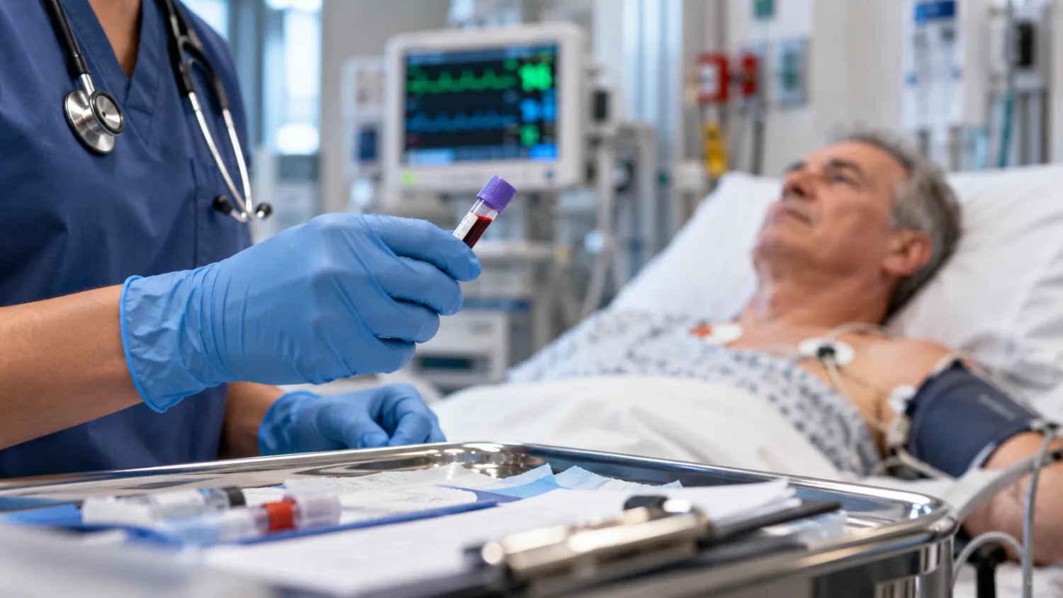
Blood tests after a low result
Tests may be arranged to look for medical factors contributing to bone loss. Depending on the circumstances, these can include:
- calcium, phosphate and alkaline phosphatase;
- vitamin D;
- kidney and liver function;
- thyroid tests;
- full blood count;
- coeliac screening;
- parathyroid hormone;
- testosterone or other hormone tests.
Our guides to calcium and bone-profile blood tests and vitamin D blood-test results explain two of the most common parts of this assessment.
Bone-strengthening medicines
Medicines may be recommended when fracture risk is sufficiently high. Options include:
- oral bisphosphonates such as alendronic acid or risedronate;
- intravenous zoledronic acid;
- denosumab;
- raloxifene in selected postmenopausal women;
- hormone replacement therapy in suitable patients;
- bone-forming medicines for some people at very high risk.
The best treatment depends on age, fracture history, kidney function, other medical conditions, pregnancy potential and whether tablets can be taken correctly.
A low T-score does not mean everyone receives the same medicine. The potential benefit must be weighed against risks and practical considerations.
Calcium and vitamin D
Calcium and vitamin D are important for bone health, but supplements are not a complete treatment for established osteoporosis when fracture risk is high.
Some people can obtain enough calcium from food. Vitamin D supplementation may be recommended when intake, sun exposure or blood levels are inadequate.
Excessive calcium supplementation is not automatically beneficial and may be unsuitable for some people with kidney stones, high blood calcium or certain medical conditions.
Exercise
Regular weight-bearing and resistance exercise supports bone and muscle health. Balance training can also reduce falls.
Appropriate activities may include:
- brisk walking;
- stair climbing;
- resistance bands or weights;
- body-weight exercises;
- balance and posture exercises.
People with spinal fractures, severe osteoporosis or significant pain should seek individual advice before starting high-impact exercise or repeated forward-bending movements.
Can DEXA results be inaccurate or misleading?
DEXA is the standard clinical method for measuring bone density, but no test is perfect. Technical and biological factors can affect the result.
Arthritis and spinal degeneration
Osteoarthritis, bony spurs and calcification can make the lumbar spine appear denser than it really is. This is particularly relevant in older adults.
The reporting clinician may exclude individual vertebrae that appear unreliable or place greater weight on the hip result.
Previous fractures
A compressed or previously fractured vertebra can alter the spinal measurement. A very dense-looking vertebra beside less dense ones may need to be excluded from analysis.
Metal implants
Hip replacements, spinal metalwork and other implants interfere with X-ray absorption. The opposite hip, forearm or another valid site may be used.
Body size and positioning
Very low or high body weight, inability to remain still and inconsistent positioning can affect precision.
A small difference between two scans may reflect measurement variation rather than a true biological change.
Different scanners
Results from different manufacturers or departments are not always directly comparable. Repeat monitoring is ideally performed on the same machine or within the same service.
A normal scan after a fracture
A normal or near-normal T-score does not prove that a fracture was unrelated to bone fragility. Bone quality, falls, certain medical conditions and factors not captured by density may still be relevant.
Osteoporosis without a very low T-score
Some people are treated as having clinically important osteoporosis because of fragility fractures or very high calculated risk, even though their lowest T-score is above −2.5.
The scan supports the clinical assessment; it does not replace it.
How often should a DEXA scan be repeated?
There is no single correct interval for everyone. The timing depends on:
- the original result;
- age and fracture risk;
- whether treatment has started;
- the medicine being used;
- new fractures;
- ongoing steroid use;
- medical conditions that may cause rapid bone loss;
- whether a repeat result would change management.
Bone density changes slowly, and repeating a scan too soon may show little more than normal measurement variation.
Some people may be rescanned after a few years, while others do not need routine repeat testing. Treatment decisions may be reviewed using fracture history, medicine adherence, side effects and changing health even when no new scan is arranged.
Does treatment have to increase the T-score?
Not necessarily. A successful treatment may stabilise bone density and reduce fracture risk even when the T-score changes only slightly.
The main purpose of treatment is to prevent fractures, not simply to improve a number on a report.
What if the result becomes worse?
A decline may lead clinicians to review:
- whether the same machine and sites were used;
- whether the change exceeds expected measurement variation;
- whether medicine has been taken correctly;
- whether an underlying condition has developed;
- whether steroid exposure has changed;
- whether a different treatment is needed.
Do not stop osteoporosis medicine solely because one result appears worse. Some treatments, particularly denosumab, require a carefully planned transition if they are discontinued.
Frequently asked questions about DEXA scans
Is a DEXA scan painful?
No. The scan is non-invasive and should not hurt. You need to lie still, and the hip may be gently positioned, which can be uncomfortable for someone with arthritis or a recent injury.
How long does a DEXA scan take?
The scan itself is often completed within about 10 to 20 minutes. The full appointment may take longer because of registration, questions, height and weight measurements and positioning.
Do I need to undress?
You may be able to remain in your own clothing if it contains no metal. Clothes with zips, buckles, hooks or underwires may need to be removed, and you may be given a gown.
Can I eat before the scan?
Usually yes. Fasting is not normally required. Follow the instructions from the scanning department, particularly regarding calcium supplements.
Can I take my usual medicines?
Usually yes. Continue prescribed medicines unless the appointment letter or clinical team instructs otherwise.
Is DEXA radiation dangerous?
DEXA uses a very low dose of ionising radiation, considerably lower than many conventional X-ray and CT examinations. The expected clinical benefit normally outweighs the small radiation risk when the scan is appropriately requested.
Can I have a DEXA scan while pregnant?
It is generally postponed during pregnancy because it uses X-rays. Tell the department if you are or could be pregnant so the clinical team can review the need and timing.
What is a good DEXA score?
For the T-score, −1.0 or above is generally considered within the normal range. However, a “good” result does not eliminate fracture risk, and a lower result does not predict that a fracture will definitely happen.
Is −2.5 worse than −2.0?
Yes. More negative T-scores represent lower measured bone density. A score of −2.5 is the conventional threshold for the osteoporosis range in postmenopausal women and men aged 50 or over.
What is the difference between osteopenia and osteoporosis?
Osteopenia describes a T-score between −1.0 and −2.5. Osteoporosis is conventionally defined by a T-score of −2.5 or lower. Overall fracture risk and previous fractures remain important regardless of the label.
Can I have osteoporosis with no symptoms?
Yes. Osteoporosis itself usually does not cause pain until a bone fractures. Spinal fractures may sometimes occur without an obvious injury and can cause height loss, a curved spine or persistent back pain.
Can a DEXA scan detect bone cancer?
No. It is not designed to diagnose bone cancer, myeloma or bone metastases. Other tests such as blood tests, X-rays, CT, MRI or nuclear-medicine scans are used when cancer is suspected.
Can DEXA diagnose arthritis?
No. It measures bone density rather than diagnosing joint disease. Arthritis can sometimes affect the accuracy of spinal measurements.
Can DEXA show vitamin D deficiency?
No. Vitamin D deficiency is assessed through history and, where appropriate, a blood test. Long-standing severe deficiency can affect bone health, but the scan cannot determine the vitamin D level.
Can I request a DEXA scan from my GP?
You can discuss your risk factors, previous fractures, medicines and family history with a GP. The GP may first complete a fracture-risk assessment and then decide whether a DEXA scan would change your care.
Should everyone over 50 have a DEXA scan?
No. Age is important, but routine scanning of every person over 50 is not generally recommended. The decision is based on fracture risk and whether the result would influence treatment.
What happens if my result shows osteopenia?
The clinician will consider the score alongside your age, fractures and other risk factors. You may receive lifestyle advice, investigation for underlying causes, follow-up or treatment if your overall fracture risk is high.
What happens if my result shows osteoporosis?
You may be offered blood tests, bone-health advice and a medicine to reduce fracture risk. The exact plan depends on your fracture history, health conditions and suitability for different treatments.
Can osteoporosis improve?
Bone density may stabilise or improve with appropriate treatment, nutrition and exercise. More importantly, effective treatment can substantially reduce the likelihood of fractures even when the T-score does not return to the normal range.