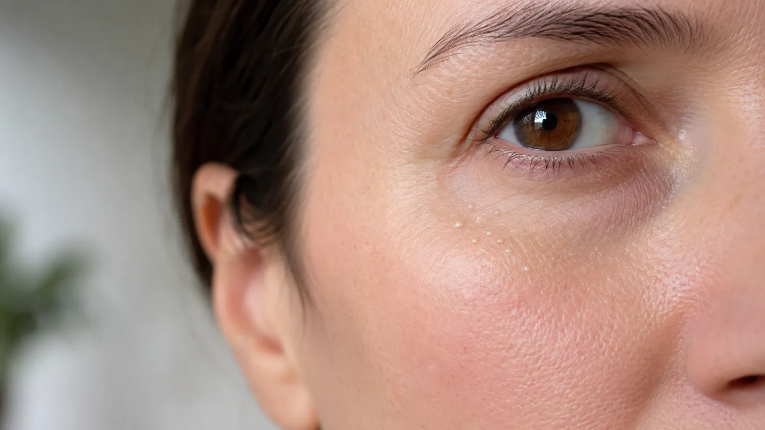
Milia are tiny white or cream-coloured bumps that form when keratin becomes trapped just beneath the surface of the skin. They are particularly common around the eyes, cheeks, forehead and nose, although they can appear almost anywhere.
Because milia resemble whiteheads, people often assume that they are a form of acne and try to squeeze them. In reality, a milium is a small enclosed cyst rather than an open or blocked pore. It usually has no natural opening through which its contents can be pushed, which is why squeezing rarely works and often leaves the skin red, bruised or scarred.
Milia are harmless and do not need treatment for medical reasons. In babies they usually disappear naturally, while in adults they may clear within weeks or months or remain unchanged for much longer. When removal is wanted, the safest option is usually professional extraction by someone appropriately trained to diagnose and treat skin lesions.
Do not puncture or cut milia at home, particularly around the eyelids. Needles, lancets, comedone tools and other sharp instruments can cause infection, bleeding, scarring or accidental injury to the eye.
Arrange a professional assessment if a supposed milium is painful, red, bleeding, crusting, rapidly enlarging, affecting vision, repeatedly returning in one place or looking different from the surrounding bumps. Not every small white or yellow eyelid lesion is milia.
What are milia?
A single bump is called a milium. The plural is milia.
Each milium is a very small cyst containing keratin, a tough structural protein found in the outer layer of skin, hair and nails. The keratin becomes enclosed beneath a thin covering of skin, creating a firm pearl-like bump.
Most milia measure only about 1 to 2 millimetres. They tend to be:
- white, ivory or pale yellow;
- round and clearly defined;
- firm rather than soft;
- smooth on the surface;
- painless;
- not itchy or inflamed;
- difficult or impossible to squeeze.
On darker skin tones, milia may appear white, cream or slightly yellow and can be particularly noticeable because of the contrast with the surrounding skin. They do not usually leave pigmentation or scars unless they are picked, squeezed or treated too aggressively.
Why are milia sometimes called milk spots?
The name “milk spots” is commonly used for milia in newborn babies. The name does not mean they are caused by milk, breastfeeding, formula or an allergy.
They are a normal and temporary skin finding in many newborns.
Are milia filled with pus?
No. The white material inside is compacted keratin, not pus.
Pus usually forms because of infection and is made from immune cells, bacteria and inflammatory material. Milia are not normally infected and do not require antibiotics.
Are milia contagious?
No. They cannot spread from one person to another through touch, towels, cosmetics or close contact.
Several bumps may develop in the same area because the skin there is prone to trapping keratin, not because one milium has infected neighbouring skin.
What causes milia?
Milia form when keratin does not shed normally from the surface of the skin and instead becomes trapped underneath it.
Sometimes this happens without an identifiable reason. In other cases, milia develop after skin injury, inflammation or the use of products that create a heavy occlusive layer over the skin.
The cause also varies according to the type of milia.
Primary milia
Primary milia arise directly from the skin without a preceding injury or rash. They develop when keratin becomes trapped within a small structure near a hair follicle or sweat duct.
These are the familiar white bumps commonly seen:
- around the eyelids;
- beneath the eyes;
- on the upper cheeks;
- on the forehead;
- around the nose.
Primary milia can occur at any age. They are very common in newborn babies but also affect children, teenagers and adults.
Secondary milia
Secondary milia develop after the skin has been damaged or inflamed. As the skin heals, keratin can become trapped beneath the newly formed surface.
Possible triggers include:
- burns;
- blistering rashes;
- abrasions and other injuries;
- prolonged use of some topical steroid creams;
- laser resurfacing;
- dermabrasion;
- chemical peels;
- radiotherapy;
- other procedures that disrupt the skin surface.
Secondary milia may appear weeks or months after the original injury has healed. They can occur in larger groups and may be more persistent than ordinary primary milia.
Sun damage
Long-term sun exposure alters the structure and shedding behaviour of the skin. Milia can develop on chronically sun-damaged areas, particularly around the eyes and upper cheeks.
Sun protection will not instantly remove established milia, but it can reduce further skin damage and may lower the likelihood of some new lesions forming.
Heavy or occlusive skincare products
Rich creams, ointments, balms and heavy make-up are sometimes blamed for every case of milia. The relationship is not that simple.
Milia can occur in people who use very little skincare, and many people use rich products without developing them. However, products that form a thick occlusive layer may contribute in someone whose skin is already prone to trapping keratin, especially around the eyes.
Potential contributors can include:
- very heavy eye creams;
- thick facial balms;
- greasy ointments;
- occlusive make-up;
- products not fully removed at night.
This does not mean that every moisturiser should be abandoned. Dry or sensitive skin still needs barrier support. A lighter, non-comedogenic formulation may simply be more comfortable for someone who repeatedly develops facial milia.
Genetic and rare skin conditions
Widespread or unusually persistent milia can occur with rare inherited or blistering skin conditions.
This is very different from having a few isolated bumps around the eyes. A person with ordinary facial milia does not normally need genetic tests or investigation for a rare disease.
Where do milia commonly appear?
Milia can form wherever suitable skin structures are present, but the face is the most familiar location.
Common areas include:
- upper and lower eyelids;
- skin beneath the eyes;
- upper cheeks;
- around the nose;
- forehead;
- chin.
They may also occur on the trunk, arms, hands or genital skin, particularly after injury or blistering.
Milia around the eyes
The skin around the eyes is thin, mobile and easily irritated. Milia here can be highly visible even when they are only a millimetre across.
The eye area is also where attempts at home extraction are most dangerous. A sudden movement, slippery tool or poorly controlled needle can damage the eyelid or surface of the eye.
Products used around the eyes must be selected carefully because acids and retinoids can cause irritation, swelling and dry-eye symptoms if they migrate into the eye.
Milia on the eyelid margin
A white bump directly on the eyelid margin may be milia, but other possibilities include:
- a blocked meibomian gland;
- a small chalazion;
- a stye;
- a cyst of an eyelid sweat gland;
- a skin tag or papilloma;
- another benign eyelid lesion.
Do not apply strong skincare acids or attempt extraction directly along the eyelash line.
Our guide to eyelid bumps, chalazia and styes explains how painful and deeper eyelid lumps differ from superficial milia.
Milia inside the mouth
Newborn babies can develop similar keratin-filled cysts on the gums or roof of the mouth. These are sometimes called Epstein pearls or Bohn nodules, depending on their location.
They are harmless and usually disappear without treatment.
How can you tell milia from whiteheads and other bumps?
The main clue is not simply that the bump is white. Its texture, position, behaviour and surrounding skin all matter.
Milia versus whiteheads
A whitehead is a closed acne comedone. Oil and dead skin cells block a pore, creating a pale or flesh-coloured bump.
Whiteheads are more likely to:
- occur alongside blackheads, spots or oily skin;
- feel softer than milia;
- develop in acne-prone areas;
- become red or inflamed;
- respond to established acne treatments.
Milia tend to look more pearly and sit as isolated, firm beads immediately beneath the skin.
Acne treatment may help when whiteheads and milia coexist, but squeezing either can increase inflammation and scarring.
See our guide to acne treatment in the UK for a fuller explanation of comedones and inflammatory spots.
Milia versus sebaceous filaments
Sebaceous filaments are normal structures that help oil travel through pores. They often appear as tiny grey, cream or yellowish dots across the nose and central face.
Unlike milia, they:
- sit inside visible pores;
- usually occur in a regular pattern;
- are flat or only slightly raised;
- refill after extraction;
- are a normal feature rather than true cysts.
Milia versus a stye
A stye is generally red, painful and tender. It forms when an eyelash follicle or eyelid gland becomes infected.
Milia are usually painless, superficial and unchanged in colour apart from their white or cream centre.
Read more in our detailed guide to styes, their causes and treatment.
Milia versus a chalazion
A chalazion is a deeper eyelid lump caused by a blocked meibomian oil gland. It is usually much larger than milia and feels as though it sits within the eyelid rather than immediately beneath its surface.
A chalazion can begin tender and later become firm and painless. Milia do not usually cause general eyelid swelling.
Milia versus xanthelasma
Xanthelasma causes soft, yellowish plaques, commonly near the inner corners of the upper or lower eyelids.
Unlike milia, xanthelasma tends to be:
- flatter;
- broader;
- yellow rather than pearly white;
- soft rather than firm;
- made from cholesterol-containing cells rather than keratin.
Xanthelasma can occur with normal cholesterol, although a clinician may recommend checking cholesterol and other cardiovascular risk factors.
Milia versus syringomas
Syringomas are benign growths arising from sweat ducts. They commonly appear as multiple small flesh-coloured or slightly yellow bumps beneath the eyes.
They may look similar to milia, but they tend to:
- match the surrounding skin more closely;
- occur symmetrically beneath both eyes;
- be slightly flatter;
- remain firmly attached rather than containing an easily extracted keratin pearl.
A dermatologist may need to distinguish the two before cosmetic treatment.
Milia versus sebaceous hyperplasia
Sebaceous hyperplasia causes enlarged oil glands, usually on the forehead or cheeks of middle-aged or older adults.
The bumps are commonly yellowish or skin coloured and may have a small central dip. They are generally larger and softer-looking than milia.
Will milia disappear on their own?
Often, yes. The skin continues renewing itself, and some milia gradually move towards the surface before releasing their contents naturally.
There is no reliable timetable. One milium may disappear within weeks, while another remains for months or years.
They are more likely to clear naturally when:
- they are small and superficial;
- they occur in a baby or young child;
- the skin has not been repeatedly picked or damaged;
- the original trigger has resolved.
Persistent milia are not necessarily unhealthy. A bump that remains unchanged for a long time may simply be enclosed too deeply to escape through normal skin shedding.
Should you leave them alone?
Leaving milia alone is the safest choice when they are correctly identified and are not bothersome.
Treatment is mainly cosmetic. There is no medical benefit in removing every tiny bump, and each procedure introduces some risk of irritation, infection, pigmentation or scarring.
Can skincare make them disappear?
Skincare may help the surface of the skin shed more evenly and can reduce the formation of some new milia. It is less reliable for removing a mature cyst that already has a complete covering.
A product can improve the surrounding texture without making every established bump disappear.
This distinction is important because repeatedly escalating to stronger acids or abrasive treatments may damage the skin while the milium remains unchanged.
How can milia be managed safely at home?
Home care should focus on supporting normal skin turnover and avoiding further blockage or injury. It should not involve cutting, piercing or forcibly extracting the bumps.
Use gentle cleansing
Wash the area with a mild cleanser that removes make-up, sunscreen and surface oil without leaving the skin tight or irritated.
Scrubbing harder does not clean out a milium because its contents are sealed beneath the skin. Excessive washing can damage the skin barrier and produce more redness and roughness.
Remove make-up before sleeping
Thorough but gentle removal is particularly useful around the eyes, where long-wear concealer, foundation and heavy eye products can accumulate.
A cleanser designed for the eye area may be preferable to aggressive rubbing with wipes.
Review very heavy products
When milia repeatedly develop in an area covered by a rich cream or balm, consider changing to a lighter formulation for several weeks.
Look for products described as:
- lightweight;
- non-comedogenic;
- oil free, where appropriate;
- suitable for sensitive skin;
- safe for use around the eyes.
“Non-comedogenic” does not guarantee that a product can never contribute to milia, but it can be a reasonable starting point.
Use sunscreen
Daily broad-spectrum sunscreen helps protect against cumulative sun damage and supports the results of treatments such as retinoids or chemical exfoliants.
Choose a formulation that feels comfortable and does not heavily coat the eye area. Sunscreen should not be abandoned because of milia; ultraviolet damage has much more important long-term consequences.
Consider a gentle chemical exfoliant
Products containing alpha hydroxy acids, such as glycolic or lactic acid, can encourage shedding of the outer skin layer.
Salicylic acid is oil soluble and may help when milia coexist with acne or blocked pores.
These products may improve fine superficial bumps and reduce new lesions, but they do not guarantee removal of mature milia.
Begin cautiously:
- choose a low-strength product;
- use it once or twice weekly at first;
- avoid broken or irritated skin;
- do not apply it directly along the eyelash line;
- stop if burning, swelling or persistent redness develops.
Using several acids, scrubs and retinoids together is more likely to cause dermatitis than faster improvement.
Topical retinoids
Retinoids such as adapalene or prescription tretinoin increase skin-cell turnover and can help some people who repeatedly develop milia, particularly when acne or sun damage is also present.
They work gradually and are more useful for prevention and general texture than for instantly emptying individual cysts.
Retinoids can cause:
- dryness;
- peeling;
- burning;
- redness;
- temporary worsening of irritation;
- increased sensitivity to sunlight.
They should not be applied too close to the eyelids unless a clinician has advised exactly how to use them. The product can migrate and cause swollen, irritated eyelids or worsen dry eyes.
Topical retinoids are generally avoided during pregnancy. Someone who is pregnant, trying to conceive or breastfeeding should seek professional advice before using medicated skincare.
Do not use harsh facial scrubs
Granular scrubs cannot open an enclosed keratin cyst. They can create microscopic skin injury and make the area look more inflamed.
If physical exfoliation is used at all, it should be gentle and infrequent. Chemical exfoliation is usually easier to control, although it can also irritate when overused.
Do not squeeze milia
Squeezing is ineffective because the cyst has no open pore. The pressure is instead absorbed by the surrounding skin.
This can cause:
- bruising;
- broken capillaries;
- inflammation;
- post-inflammatory dark marks;
- infection;
- permanent scarring.
The risk is especially high in delicate under-eye skin.
How are milia removed professionally?
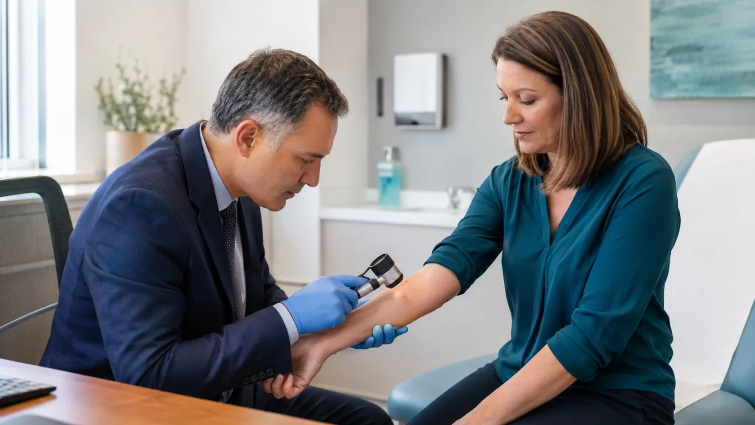
When milia are persistent or cosmetically bothersome, a dermatologist or another appropriately trained professional can confirm the diagnosis and discuss removal.
The right method depends on the number, depth, location and surrounding skin.
Manual extraction
Manual extraction is often the simplest treatment for a small number of superficial milia.
The professional cleans the skin and creates a very small opening in the surface using a sterile needle or fine blade. The keratin pearl is then removed with gentle pressure or a specialised instrument.
The key difference from squeezing at home is that the cyst covering is opened in a controlled, sterile way before pressure is applied.
After extraction, the area may show:
- a tiny red mark;
- minor swelling;
- a pinpoint scab;
- temporary tenderness.
It usually heals quickly when left clean and untouched.
Electrocautery or diathermy
Electrical energy can create a tiny controlled opening or destroy the cyst.
This may be useful for multiple lesions, but it carries risks including burns, scarring and pigmentation changes. Treatment should be adapted carefully for darker skin tones, where post-inflammatory hyperpigmentation may be more noticeable.
Laser treatment
Some dermatology and cosmetic clinics use laser treatment for extensive or resistant milia.
Laser is rarely necessary for a few ordinary superficial bumps. It may be considered when:
- there are many lesions;
- milia are associated with scarring or sun damage;
- other methods have not worked;
- another skin concern is being treated at the same time.
Laser treatment has costs and potential complications, including redness, swelling, pigmentation changes and scarring.
Cryotherapy
Freezing can destroy some superficial lesions, but it is not normally the first choice for tiny facial milia because surrounding skin can also be damaged.
Temporary blistering, lighter or darker pigmentation and scarring are possible.
Professional chemical peels
Chemical peels may improve widespread rough texture and help prevent new milia, particularly when sun damage or acne is also present.
A peel is not always the most efficient way to remove several individual mature cysts. Direct extraction may be more precise.
Will milia return after removal?
The treated cyst is unlikely to refill when its contents and covering have been adequately cleared. New milia can still form nearby because the skin remains prone to them.
Recurrence does not necessarily mean that the extraction was performed incorrectly.
Does the NHS remove milia?
Because milia are harmless and removal is usually cosmetic, treatment may not be routinely funded by the NHS.
NHS assessment may still be appropriate when:
- the diagnosis is uncertain;
- a lesion has suspicious features;
- there is significant inflammation or another medical condition;
- a child has lesions affecting vision;
- milia are associated with extensive blistering or scarring disease.
Private cosmetic treatment should be provided only after the practitioner has confirmed that the bumps are genuinely milia.
Milia in babies and children
Milia are extremely common in newborn babies. They usually appear as tiny white dots across the nose, cheeks, chin or forehead.
They do not hurt, itch or make the baby unwell. They are not caused by milk, maternal diet, formula, poor washing or infection.
Newborn milia generally disappear naturally as the skin matures and renews itself. No treatment is needed. NHS and neonatal guidance describes them as harmless and self-resolving.
How should newborn milia be cared for?
Wash the baby’s face gently with water and use mild products appropriate for newborn skin where needed.
Do not:
- squeeze the bumps;
- scrub the face;
- apply adult acne products;
- use acids or retinoids;
- attempt extraction;
- apply essential oils or strong home remedies.
The skin usually clears within the first several weeks, although some lesions can take longer.
What if the bumps are red?
Newborn acne can cause red spots, pustules and whiteheads and may be mistaken for milia. Other newborn rashes can also produce bumps.
Seek medical advice when a young baby has:
- fever;
- blisters;
- rapidly spreading redness;
- poor feeding;
- unusual sleepiness;
- breathing difficulty;
- a rash that does not fade when pressed.
These are not expected features of ordinary milia.
Persistent milia in older children
A few persistent lesions are usually harmless. Widespread, recurring or unusual milia may justify assessment by a GP or dermatologist, particularly when they occur with blistering, fragile skin, scarring or nail changes.
When should milia be checked by a professional?
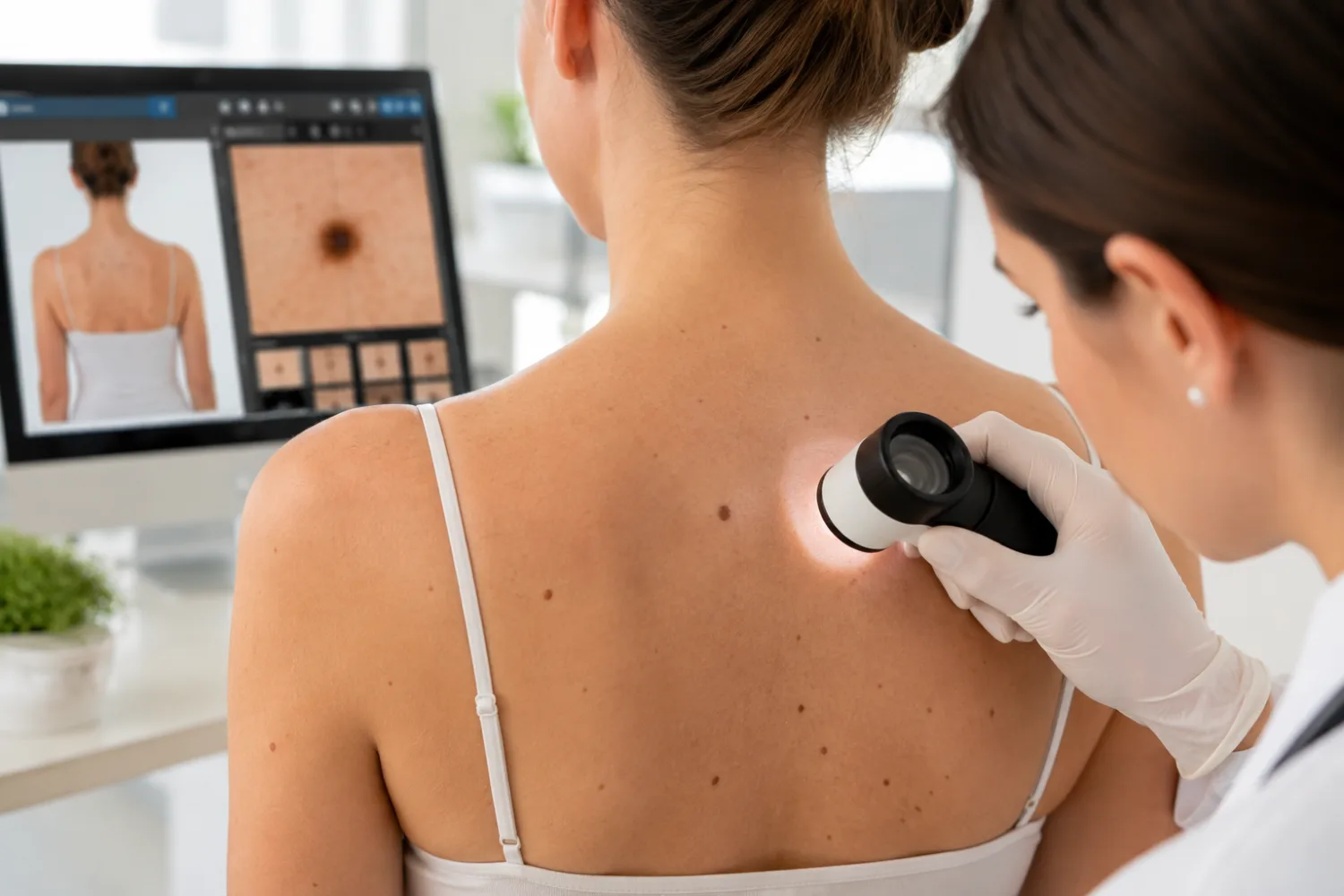
Most typical milia do not require a medical appointment. Assessment becomes useful when the diagnosis is uncertain or the lesion behaves differently from an ordinary harmless cyst.
Arrange a routine assessment if:
- you are not sure whether the bumps are milia;
- they are widespread or rapidly increasing;
- they persist despite a sensible skincare routine;
- they repeatedly form after skin injury or blistering;
- you want professional removal;
- they are very close to the eyelid margin;
- a large number developed after a cosmetic procedure;
- a child has persistent lesions affecting the eyelids.
Seek earlier advice if a bump:
- becomes painful or very tender;
- turns red and swollen;
- releases pus;
- bleeds or repeatedly crusts;
- ulcerates;
- grows quickly;
- causes eyelash loss;
- changes the shape of the eyelid;
- affects vision.
These features suggest either inflammation, infection or another diagnosis.
Urgent eye symptoms
Milia themselves should not cause:
- pain inside the eye;
- marked sensitivity to light;
- blurred or reduced vision;
- painful eye movements;
- a bulging eye;
- severe eyelid swelling;
- fever or significant illness.
Seek urgent eye care when any of these symptoms occur. Read our guide to red eyes, eye pain and when eye symptoms are urgent.
Could a persistent white bump be skin cancer?
Most small white facial bumps are benign. Some basal cell carcinomas and other skin lesions can nevertheless appear pale, pearly or skin coloured.
Warning features include:
- steady enlargement;
- bleeding with minimal contact;
- an ulcer that does not heal;
- visible small blood vessels;
- recurrent crusting;
- distortion of the eyelid margin;
- loss of eyelashes.
A changing lesion should not be repeatedly treated as milia without a confirmed diagnosis. Our guide to skin cancer checks in the UK explains when a lesion should be examined.
Frequently asked questions about milia
What are milia made of?
They contain compacted keratin, a protein found in skin, hair and nails.
Are milia a type of acne?
No. They are tiny keratin-filled cysts. They can occur alongside acne, but they are not whiteheads and do not form through exactly the same process.
Why can’t milia be squeezed?
A milium is enclosed beneath a layer of skin and normally has no open pore. Squeezing presses on the surrounding tissue without giving the keratin a way to escape.
What is the fastest way to remove milia?
For a small number of established milia, professional sterile extraction is usually the fastest and most direct method.
Can I remove milia with a needle?
You should not attempt this at home. There is a risk of infection, bleeding, scarring and accidental eye injury, particularly around the eyelids.
Do milia go away naturally?
Many do. They may clear within weeks or months, while some adult milia remain much longer.
How long do milia last?
There is no fixed duration. Newborn milia usually clear quickly, while adult lesions may last for months or occasionally years.
Does moisturiser cause milia?
Not necessarily. Heavy occlusive products may contribute in someone who is prone to them, but milia also develop without skincare products.
Should I stop using eye cream?
Not automatically. Consider switching to a lighter product if milia repeatedly appear in the exact area where a heavy eye cream is applied.
Does Vaseline cause milia?
Petrolatum does not cause milia in everyone and can be valuable for protecting very dry or damaged skin. Its highly occlusive nature may feel too heavy for some milia-prone facial areas.
Can sunscreen cause milia?
Any product may feel too heavy for an individual’s skin, but sunscreen should not simply be abandoned. Try a lighter non-comedogenic formulation while maintaining daily ultraviolet protection.
Can retinol remove milia?
Retinol and prescription retinoids may improve skin turnover and help prevent some milia. They do not guarantee that a mature enclosed cyst will disappear.
Can salicylic acid remove milia?
It may help superficial texture and blocked pores, particularly when acne is also present. It may not penetrate and remove an established keratin cyst.
Can glycolic acid remove milia?
Glycolic acid can encourage surface exfoliation and may gradually improve small superficial lesions. Strong or frequent use can irritate the skin, especially around the eyes.
Can facials remove milia?
A trained practitioner may remove confirmed superficial milia during a facial treatment. Ordinary steaming or exfoliation alone will not necessarily extract a mature cyst.
Are milia caused by poor hygiene?
No. They result from trapped keratin and can occur in clean, healthy skin.
Are milia caused by high cholesterol?
No. Xanthelasma, which causes broader yellow plaques around the eyelids, may sometimes be associated with high cholesterol. Milia contain keratin rather than cholesterol.
Are milia caused by vitamin deficiency?
Ordinary facial milia are not generally considered a sign of vitamin deficiency. Taking supplements without a diagnosed deficiency is unlikely to remove them.
Can stress cause milia?
Stress is not a direct cause. It may alter skincare habits or aggravate related skin conditions, but it does not itself create keratin cysts.
Can milia become infected?
They are not normally infected. Picking, puncturing or squeezing them can introduce bacteria and cause redness, pain or pus.
Do milia leave scars?
Untouched milia usually do not scar. Scarring is more likely after repeated squeezing, digging, burns from acids or poorly performed extraction.
Can milia return after extraction?
The extracted lesion may be gone, but new milia can develop nearby if the skin remains prone to them.
Are milia common in babies?
Yes. They are a very common and harmless newborn skin finding and usually disappear without treatment.
Should baby milia be treated?
No. Keep the skin clean and leave the bumps alone. Do not apply acne medicines, acids or adult retinoids.
Can milia occur on the lips?
Small pale bumps around the lips may have several causes, including milia and Fordyce spots. A clinician can assess persistent or uncertain lesions.
Can milia grow larger?
Ordinary milia usually remain very small. A steadily enlarging lump may be another type of cyst or skin lesion and should be examined.
Who should remove milia around the eyes?
A dermatologist, appropriately trained medical professional or experienced eye specialist should assess lesions very close to the eyelid margin. Diagnosis is particularly important before any procedure near the eye.