A private echocardiogram typically costs around £250 to £500 in the UK for a standard transthoracic scan with a written report. A package that also includes a consultation with a cardiologist may cost approximately £450 to £800 or more, depending on the clinic, location and complexity of the assessment.
An echocardiogram—often shortened to an “echo”—is an ultrasound scan of the heart. It shows the heart chambers, muscle, valves and blood flow in real time. It can help investigate breathlessness, a heart murmur, swelling, chest symptoms or an abnormal ECG, but it does not answer every possible question about the heart.
The lowest advertised price may cover only the scan and report. A cardiologist’s consultation, ECG, blood tests, stress testing and follow-up discussion can all be charged separately. Before booking, check exactly who reviews the scan, when you receive the report and what happens if it identifies an abnormality.
This guide provides general information about private echocardiogram prices and testing in the UK. New or severe chest pain, major breathing difficulty, collapse or other emergency symptoms require urgent medical help rather than a routine private scan.
How much does a private echocardiogram cost in the UK?
A standard private transthoracic echocardiogram commonly costs between £250 and £500. Prices tend to be lower at dedicated diagnostic centres and higher at private hospitals or consultant-led cardiology clinics.
Current representative examples illustrate the variation. Vista Health advertises echocardiograms from £250, including a written report with consultant cardiologist oversight. Scan.com also advertises a £250 end-to-end echocardiogram package. Nuffield Health lists prices from £350 at its Edinburgh hospital and a guide price of £399 at its Manchester Diagnostic Suite.
At the more expensive end, some London hospitals charge over £700 when the service includes cardiologist involvement, results and explanation at the appointment.
| Private heart service | Typical self-pay price |
|---|---|
| Standard transthoracic echocardiogram with report | £250–£500 |
| Echocardiogram plus cardiologist consultation | £450–£800+ |
| ECG added to the assessment | Approximately £50–£150 extra, if not included |
| Stress echocardiogram | Approximately £500–£900+ |
| Contrast or bubble echocardiogram | Often priced individually according to the indication |
| Transoesophageal echocardiogram | Usually considerably more than a standard echo |
These are broad market ranges rather than fixed tariffs. The same provider may charge different prices at different hospitals, and specialist scans can involve additional facility, medication and consultant fees.
For a wider comparison of cardiac assessment fees, see our guide to private cardiology tests, consultations and insurance cover.
What should the advertised price include?
A basic echocardiogram price should normally include the scan itself and a clinical report. It may also include review or reporting by a cardiac physiologist, echocardiographer or consultant cardiologist.
However, “echocardiogram from £250” does not necessarily include a conversation with a cardiologist. In some services, you receive a written report and are expected to take it to your GP or referring specialist.
Before paying, ask whether the fee includes:
- the complete transthoracic scan, including Doppler measurements;
- a written clinical report;
- consultant cardiologist review or oversight;
- an explanation of the findings;
- sending the report to your GP or specialist;
- images or digital access to the scan;
- an ECG performed at the same appointment;
- contrast if ordinary images are unclear;
- a follow-up appointment if the result is abnormal;
- the provider’s cancellation and rescheduling terms.
Ask who takes responsibility for acting on an important result. A scan without an appropriate clinical review may leave you with measurements but no clear plan.
A cardiologist consultation commonly adds approximately £200 to £350, although charges vary widely. Blood tests, ambulatory heart monitoring, cardiac MRI or CT scans would normally be separate.
Our guide to private MRI, CT and ultrasound scan costs explains how reporting and consultant fees affect the final price of diagnostic imaging.
What is an echocardiogram?
An echocardiogram uses high-frequency sound waves to create moving images of the heart. It does not use X-rays or ionising radiation.
The standard test is called a transthoracic echocardiogram, or TTE. “Transthoracic” means the heart is examined through the chest wall.
A handheld ultrasound probe is moved across different areas of the chest. Gel placed between the probe and skin helps the sound waves travel effectively.
The returning sound waves are converted into images of the beating heart. Doppler ultrasound can also measure the direction and speed of blood moving through the chambers and valves.
The NHS describes an echocardiogram as an ultrasound test used to create images of the heart and assess how well it is working. The standard scan is usually performed by a specialist healthcare professional known as a sonographer or echocardiographer. You can read the NHS overview of what happens during an echocardiogram.
What does an echocardiogram show?
An echocardiogram provides information about both the structure and function of the heart. The report brings several measurements and observations together rather than giving one simple “positive” or “negative” result.
Heart chamber size
The scan measures the left and right atria and ventricles. Enlargement can occur with valve disease, high blood pressure, abnormal heart rhythms, cardiomyopathy and other conditions.
A mildly enlarged measurement does not establish the cause by itself. It needs to be interpreted alongside body size, symptoms, blood pressure, ECG findings and medical history.
Heart muscle thickness
An echo can show whether the ventricular walls are unusually thick. Long-term high blood pressure can make the left ventricle work harder and become thickened, a finding known as left ventricular hypertrophy.
Thickening can also have other causes, including inherited heart muscle conditions. The pattern and degree therefore matter.
If blood pressure is part of the concern, see our guides to high blood pressure and monitoring blood pressure at home.
Pumping function
The scan assesses how well the heart muscle contracts. One commonly reported measurement is the left ventricular ejection fraction, or LVEF.
Ejection fraction describes the proportion of blood pushed out of the left ventricle with each contraction. The British Heart Foundation explains that a normal ejection fraction is generally above approximately 50% to 55%, although laboratories and clinical contexts differ.
A reduced ejection fraction can be associated with heart failure, cardiomyopathy, previous heart-muscle damage and several other conditions. A normal ejection fraction does not completely exclude heart failure because the heart can have difficulty relaxing or filling even when the percentage pumped out remains preserved.
Ejection fraction is also an estimate rather than a perfect measurement. Small differences between two scans may reflect image quality, technique or normal measurement variation rather than a meaningful deterioration.
Heart-valve function
An echocardiogram shows the four main heart valves and how blood moves through them. It can identify valves that are narrowed, known as stenosis, or valves that allow blood to leak backwards, known as regurgitation.
Doppler measurements help estimate the speed of blood flow, pressure gradients and severity of valve disease. Mild valve leakage is a common finding and is not always clinically important.
More significant abnormalities may require cardiology review, repeat monitoring or treatment. The decision depends on the affected valve, severity, symptoms, heart size and pumping function.
Heart movement and regional abnormalities
The echocardiographer checks whether different areas of the heart wall contract normally. A section moving less than expected may suggest previous heart-muscle injury or reduced blood supply, although other conditions can cause similar findings.
A resting echocardiogram cannot reliably exclude narrowing in the coronary arteries. When coronary disease is suspected, a stress echo, CT coronary angiogram or another test may be more appropriate.
Fluid around the heart
The scan can detect fluid in the sac surrounding the heart, known as a pericardial effusion. Small amounts may be monitored, while larger or haemodynamically important collections require urgent specialist assessment.
Pressure and blood-flow estimates
Doppler findings can help estimate pressures inside parts of the heart and pulmonary circulation. These estimates may raise concern about pulmonary hypertension, but confirmation and assessment of the cause can require additional testing.
Congenital and structural abnormalities
An echo may identify holes between heart chambers, abnormal connections or other structural differences. Some subtle defects are difficult to see on a standard scan and may require a bubble study, transoesophageal echo, cardiac MRI or other specialist imaging.
Why might a doctor recommend an echocardiogram?
An echo may be requested after symptoms, examination findings or another test suggest that the heart’s structure or pumping function should be assessed.
Common reasons include:
- a heart murmur heard through a stethoscope;
- unexplained breathlessness;
- ankle or leg swelling;
- suspected heart failure;
- an abnormal ECG;
- persistent palpitations with other concerning findings;
- high blood pressure with suspected heart-muscle changes;
- known or suspected heart-valve disease;
- possible cardiomyopathy;
- monitoring an existing heart condition;
- assessment after certain cancer treatments;
- a family history of particular inherited cardiac conditions.
An echo is not automatically the correct test for every palpitation or episode of chest discomfort. Palpitations are often investigated initially with an ECG or longer heart-rhythm monitor because an echo shows structure and function rather than recording intermittent rhythms over hours or days.
Our guides to heart palpitations and ECG results explain how these symptoms and tests fit together.
What cannot an echocardiogram show?
An echocardiogram is an important heart test, but a normal result does not mean that every possible heart problem has been excluded.
A standard resting echo does not reliably:
- show whether the coronary arteries are narrowed;
- rule out angina or an impending heart attack;
- record intermittent abnormal rhythms over an extended period;
- identify every cause of chest pain or breathlessness;
- replace blood tests for heart-muscle injury;
- provide the same tissue detail as a cardiac MRI;
- exclude all small structural defects;
- determine cardiovascular risk from cholesterol, smoking and other factors.
An ECG records the electrical activity of the heart. An echocardiogram creates moving ultrasound images of its structure and function. The two tests provide different information and are often used together.
A CT coronary angiogram is more suited to examining the coronary arteries. Cardiac MRI can provide detailed information about heart-muscle tissue, inflammation and scarring. An ambulatory ECG monitor is used when the priority is capturing intermittent rhythm disturbances.
Do not use a privately purchased normal echo as reassurance against new chest pain. Our guide to chest pain and when to seek urgent help explains the warning signs that should not wait for routine testing.
What are the different types of echocardiogram?
Most people need a standard transthoracic echo. Specialist forms are used when ordinary imaging cannot answer the clinical question.
Transthoracic echocardiogram
This is the routine non-invasive scan performed through the chest wall. It usually takes around 30 to 45 minutes and requires no sedation or recovery period.
Stress echocardiogram
A stress echo compares the heart at rest and while it is working harder. Stress may be created by exercise or medication.
The test looks for changes in heart-wall movement and function that become apparent under stress. It may be used when reduced blood supply to the heart muscle or the effect of valve disease during exertion is being investigated.
A stress echo needs more clinical supervision than a resting scan, so it costs more and should be selected for a specific medical indication.
Transoesophageal echocardiogram
A transoesophageal echocardiogram, or TOE, uses a specialised probe passed down the oesophagus. Because the oesophagus lies close to the heart, this can provide clearer images of certain valves and structures.
The throat is numbed and sedation may be used. You will need preparation, monitoring and recovery instructions, and you may need someone to accompany you home. This is not equivalent to a standard walk-in ultrasound appointment.
Contrast echocardiogram
A contrast agent may be injected into a vein when ordinary images do not show the chamber borders clearly enough. The contrast helps the clinician assess the heart more accurately.
The provider should explain the reason, possible reactions and whether contrast is included in the quoted fee.
Bubble echocardiogram
A bubble study uses agitated saline injected through a vein while ultrasound images are taken. It can help investigate whether blood is passing through an abnormal connection between the right and left sides of the heart.
This is sometimes requested following particular types of stroke or when a patent foramen ovale is suspected. Finding a PFO does not automatically mean that treatment or closure is required.
What happens during a standard echo?
You can normally eat, drink and take regular medication as usual before a transthoracic echocardiogram unless the clinic gives different instructions.
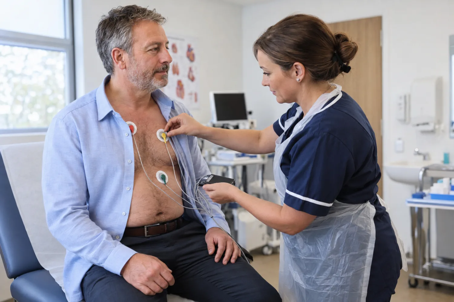
At the appointment, you will generally be asked to remove clothing from the upper body and put on a gown. Small adhesive electrodes may be placed on the chest to monitor the heartbeat and time the ultrasound images.
You will usually lie on your left side. The echocardiographer applies gel and presses the probe against several parts of the chest. You may be asked to change position, breathe in a particular way or briefly hold your breath.
The examination may involve firm pressure between the ribs to obtain a clear view. This can be uncomfortable, especially when the chest is tender, but a standard echo should not normally be painful.
Obtaining useful images can be more difficult in some people because of body shape, lung disease, previous surgery or the position of the heart. This does not necessarily mean that anything is wrong. Contrast or a different imaging test may occasionally be recommended.
The scan generally takes 20 to 45 minutes. You can normally leave immediately and return to usual activities.
How are echocardiogram results reported?
The person performing the scan may not provide a complete interpretation during the appointment. Images and measurements often need formal review before the report is finalised.
A report may include:
- left and right ventricular size and function;
- ejection fraction or a qualitative assessment of pumping function;
- heart-wall thickness;
- atrial size;
- valve appearance and function;
- Doppler blood-flow measurements;
- estimated pressures;
- the appearance of the aorta where visible;
- whether fluid is present around the heart;
- image-quality limitations;
- a conclusion and any suggested follow-up.
Some diagnostic centres return a report within one or two working days, while others take several days. Same-day results are more likely when a cardiologist consultation is included, but this should be confirmed before booking.
Terms such as “mild regurgitation”, “impaired relaxation” or “borderline chamber enlargement” can sound alarming without context. Some findings are common, age-related or not clinically important. Interpretation should consider symptoms and other tests rather than one isolated phrase.
Ask these questions after an abnormal result:
- What exactly was found?
- How severe is it?
- Could it explain my symptoms?
- Do I need treatment or monitoring?
- When should the echo be repeated?
- Do I need an ECG, blood tests, CT or cardiac MRI?
- Which symptoms should prompt urgent help?
Can you book an echocardiogram without a referral?
Some UK diagnostic centres accept self-referrals for a standard echocardiogram. Others require a referral from a GP, cardiologist or another clinician.
Self-referral may provide faster access, but it does not remove the need for clinical reasoning. The important question is not simply whether you can buy the scan, but whether an echo is the right investigation for your symptoms.
For example, intermittent palpitations may require prolonged ECG monitoring. Suspected angina may need coronary imaging or functional testing. Severe breathlessness can have cardiac, respiratory, vascular or other causes.
A private GP or cardiologist can review the symptoms and select the most useful test. If you already have an NHS referral, paying privately for the scan does not necessarily remove you from NHS care.
Our guides to how hospital referrals work and same-day private GP appointment costs explain the available routes.
Will private health insurance cover an echocardiogram?
Private medical insurance may cover an echocardiogram when it is medically necessary, requested by an eligible clinician and performed by an approved provider.
Insurers commonly require:
- a GP or specialist referral;
- pre-authorisation before the scan;
- use of a hospital or clinician recognised by the insurer;
- the condition not to be excluded as pre-existing or chronic;
- payment of the policy excess.
Do not book and pay first on the assumption that the insurer will reimburse you. Ask whether the consultation, scan, ECG and follow-up are all covered under the same authorisation.
Some policies cover diagnosis but limit ongoing monitoring of a chronic heart condition. If the echo is part of routine surveillance rather than investigation of a new symptom, check the wording carefully.
Our guides to what private health insurance covers and making a private health insurance claim explain the process in more detail.
When should you seek urgent medical help instead?
A private echocardiogram is a planned diagnostic test, not an emergency assessment. Do not wait for a routine appointment if symptoms could indicate a heart attack, pulmonary embolism or another acute condition.
Call 999 for symptoms such as:
- sudden or severe chest pressure, tightness or heaviness;
- chest discomfort spreading to the arm, jaw, back or stomach;
- major breathing difficulty;
- collapse or loss of consciousness;
- chest symptoms with sweating, nausea or a sense of impending doom;
- a very fast or abnormal heartbeat with collapse, severe breathlessness or chest pain.
Seek prompt medical advice for new or worsening breathlessness, fainting, increasing leg swelling or reduced ability to exercise. These symptoms need a clinical assessment even if you intend to arrange an echo privately.
A normal echo does not rule out a future heart attack. Long-term prevention also involves blood pressure, cholesterol, diabetes, smoking, weight, activity and family history. See our complete guide to cardiovascular risk and prevention.
Frequently asked questions
How much is a private echocardiogram in the UK?
A standard transthoracic echocardiogram with a written report commonly costs £250 to £500. A package including a cardiologist consultation may cost around £450 to £800 or more.
Is an echocardiogram the same as an ECG?
No. An ECG records the heart’s electrical activity using electrodes on the skin. An echocardiogram uses ultrasound to produce moving images of the heart’s chambers, muscle, valves and blood flow.
Does an echocardiogram show blocked arteries?
A standard resting echo does not directly show most coronary-artery narrowing and cannot reliably exclude coronary heart disease. A stress test, CT coronary angiogram or other investigation may be required.
Can an echo detect heart failure?
It can provide important evidence about pumping function, relaxation, chamber size, valves and pressures. Heart failure is diagnosed using the complete clinical picture, not the scan alone.
What is ejection fraction?
Ejection fraction estimates the percentage of blood pushed out of the left ventricle with each contraction. A normal value is commonly above approximately 50% to 55%, but the result must be interpreted with other measurements and symptoms.
Can you have heart failure with a normal ejection fraction?
Yes. Some people have heart failure with preserved ejection fraction, where contraction remains relatively normal but the ventricle is stiff or does not fill effectively.
Does an echo show previous heart attacks?
It may show areas of heart muscle that move abnormally or reduced overall pumping function, which can occur after a heart attack. A normal resting echo cannot exclude all previous damage or coronary disease.
Is an echocardiogram painful?
A standard transthoracic echo is non-invasive and normally painless. Pressure from the probe can feel uncomfortable between the ribs or over a tender area of the chest.
Does an echocardiogram use radiation?
No. It uses ultrasound sound waves and does not expose you to ionising radiation.
How long does a private echocardiogram take?
A standard scan usually takes approximately 20 to 45 minutes. Extra time may be needed when images are difficult to obtain or when contrast is used.
Do I need to fast before an echo?
Not for a routine transthoracic echo. A transoesophageal or stress echo may have specific food, drink and medication instructions, which the clinic should provide in advance.
Can I drive after an echocardiogram?
You can normally drive immediately after a standard transthoracic scan. You should not drive after sedation for a transoesophageal echo and will need to follow the provider’s discharge instructions.
How quickly will I receive the report?
Some providers return reports within one or two working days, while others take several days. A same-day explanation may be available when the scan is part of a cardiologist consultation.
What happens if the private echo is abnormal?
The report may recommend GP or cardiology review, repeat monitoring or another test. Ask the provider who will explain the result and arrange an appropriate clinical plan before booking the scan.
Can I take private echo results to my NHS GP?
Yes. Your NHS GP can review the report and make an independent decision about NHS treatment or referral. A private result does not automatically guarantee a particular NHS test or specialist appointment.
Is a private echo worth paying for?
It may be worthwhile when an echo is clinically appropriate and quicker access would influence diagnosis, reassurance or treatment. Its value is lower when the wrong test is purchased or the price includes no adequate interpretation or follow-up.