A CT scan is one of the most commonly used hospital imaging tests. It can give doctors fast, detailed pictures of the inside of the body and is especially useful in emergencies, cancer care, chest and abdominal symptoms, injuries, kidney stones, infections, bleeding and complex conditions where a standard X-ray or ultrasound is not enough.
You may be sent for a CT scan after symptoms, blood tests, an X-ray, an ultrasound, an MRI, a hospital admission, a cancer referral or an emergency assessment. Some people also have CT scans privately, often after specialist advice or as part of a diagnostic pathway.
CT scans are quick, painless and widely used. However, they do involve X-ray radiation, and some CT scans use contrast dye, which has its own small risks. This does not mean CT should be avoided when it is needed. It means the scan should be requested for a clear reason, performed safely, and interpreted in context.
This guide explains what a CT scan is, what it can show, when it is needed, how it differs from MRI and ultrasound, how to prepare, what contrast dye is, what the radiation risk means, when results come back, and what to ask your doctor.
Quick answer: A CT scan uses X-rays and a computer to create detailed cross-sectional images of the body. It is fast and especially useful for emergencies, chest problems, abdominal pain, injuries, bleeding, kidney stones, infections and cancer assessment. CT differs from MRI because CT uses radiation and is usually quicker, while MRI uses magnets and is often better for soft tissues such as brain, spine, joints and pelvis.
What is a CT scan?
CT stands for computed tomography. A CT scanner uses X-rays taken from different angles around the body. A computer then combines these images to create detailed slices, or cross-sectional pictures, of the area being scanned.
Instead of one flat picture like a standard X-ray, CT gives doctors a much more detailed view. It can show bones, organs, blood vessels, soft tissues, air spaces, fluid, bleeding, masses and infection. The images can also be reconstructed in different planes or 3D views when needed.
The NHS explains that CT scans use X-rays and a computer to create detailed images of the inside of the body. They are carried out in hospital by specially trained radiographers and are usually quick and painless. NHS CT scan guidance.
How does a CT scan work?
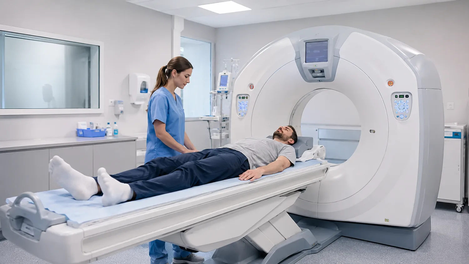
During a CT scan, you lie on a moving table that passes through a doughnut-shaped scanner. The scanner rotates around the body and takes X-ray images from many angles. These are processed by a computer to create detailed pictures.
The scan itself may only take a few minutes, although the whole appointment may take longer because of preparation, safety checks, contrast dye, cannula placement or observation afterwards.
CT is particularly useful because it is fast. In emergency medicine, speed matters. A CT scan can quickly help doctors look for bleeding, stroke changes, internal injuries, blood clots, bowel obstruction, kidney stones, severe infection or cancer complications.
What can a CT scan show?
CT can show many parts of the body, depending on the area scanned and whether contrast dye is used.
A CT scan can help assess:
- Brain: bleeding, stroke changes, tumours, swelling, skull fractures and head injury.
- Chest: lung infection, lung cancer, blood clots, fluid, collapsed lung and some heart or vessel problems.
- Abdomen and pelvis: bowel obstruction, appendicitis, diverticulitis, kidney stones, pancreatitis, liver problems, masses, infection and internal bleeding.
- Bones and trauma: fractures, complex injuries, spinal injuries and surgical planning.
- Blood vessels: aneurysms, narrowing, blockages, pulmonary embolism and bleeding when CT angiography is used.
- Cancer care: diagnosis, staging, treatment planning, response monitoring and follow-up.
- Infection and inflammation: abscesses, severe infections, inflammatory complications and fluid collections.
CT is not always the best test for every condition. MRI, ultrasound, X-ray, blood tests, stool tests, gastroscopy or colonoscopy may be better depending on the question. See our guides to MRI scans, abdominal ultrasound, gastroscopy and colonoscopy.
When is a CT scan needed?
A CT scan is requested when the result may help diagnose a condition, guide treatment, plan surgery, monitor disease or clarify something found on another test. It is often used when doctors need more detail than a standard X-ray or ultrasound can provide.
Common reasons include:
- head injury or suspected brain bleeding
- stroke assessment in some situations
- chest pain or suspected blood clot in the lung
- shortness of breath with concerning symptoms
- persistent cough with abnormal X-ray findings
- severe abdominal pain
- suspected appendicitis or diverticulitis
- suspected kidney stones
- vomiting with suspected bowel obstruction
- unexplained weight loss
- blood in urine
- suspected cancer or cancer staging
- trauma after a fall, crash or major injury
- checking infection, abscess or inflammation
- follow-up of abnormal ultrasound, MRI or blood tests
If your CT is being requested for symptoms, related guides include abdominal pain, chest pain, cough in adults, blood in stool, pain when urinating and unexplained weight loss.
CT scan of the head
A head CT is often used in emergencies because it is fast and good at showing bleeding, swelling, skull fractures and some stroke changes. It may be requested after head injury, sudden severe headache, weakness, confusion, seizures or suspected bleeding.
A head CT can show:
- bleeding in or around the brain
- skull fractures
- brain swelling
- some strokes
- large tumours or masses
- fluid build-up
- some infections or complications
MRI may be used later if more detail is needed, especially for certain neurological conditions, small lesions, multiple sclerosis, some tumours or subtle stroke changes.
CT scan of the chest
A chest CT gives detailed images of the lungs, airways, lymph nodes, chest wall, blood vessels and surrounding structures. It can show much more detail than a standard chest X-ray.
A chest CT may be requested for:
- abnormal chest X-ray findings
- suspected lung cancer
- persistent or unexplained cough
- shortness of breath
- suspected pulmonary embolism, meaning a blood clot in the lung
- pneumonia complications
- interstitial lung disease
- chest trauma
- fluid around the lungs
- cancer staging or follow-up
A CT pulmonary angiogram, often called CTPA, uses contrast dye to look for blood clots in the lungs. This is a common emergency scan when pulmonary embolism is suspected.
CT scan of the abdomen and pelvis
CT is very useful for abdominal and pelvic symptoms because it can show many organs, blood vessels, bowel loops, lymph nodes and deeper structures in one scan.
An abdominal or pelvic CT may help diagnose:
- appendicitis
- diverticulitis
- bowel obstruction
- kidney stones
- pancreatitis
- abscesses or infection
- internal bleeding
- some liver, spleen, pancreas, kidney or adrenal problems
- cancers or enlarged lymph nodes
- complications of inflammatory bowel disease
Ultrasound may be used first for gallstones, liver, kidneys or pelvic organs, while CT may be used when symptoms are severe, unclear, urgent or when more detail is needed. See diverticular disease and diverticulitis, gallstones, IBD explained and kidney blood test results.
CT scan for cancer diagnosis and monitoring
CT plays a major role in cancer care. It can help find tumours, assess whether cancer has spread, guide biopsies, plan surgery or radiotherapy, monitor response to treatment and check for recurrence.
In cancer pathways, CT may be used alongside blood tests, MRI, ultrasound, PET-CT, endoscopy, colonoscopy, mammography, biopsy or specialist scans. The choice depends on cancer type and clinical question.
CT is not a perfect cancer test. Some cancers are better seen with MRI, ultrasound, endoscopy or other tests. A CT report should always be interpreted by the clinical team alongside symptoms, examination and test results.
CT scan for kidney stones
CT is often very good at detecting kidney stones, especially stones in the ureter, the tube between kidney and bladder. In many cases, CT can show the stone size, location and whether it is causing blockage.
Ultrasound can detect some stones and kidney swelling, but CT is often more sensitive for stones. The exact test depends on age, pregnancy possibility, symptoms, severity and local protocols.
CT angiography: scanning blood vessels
CT angiography uses contrast dye to show blood vessels. It can help assess arteries and veins in different parts of the body.
It may be used for:
- pulmonary embolism
- aortic aneurysm
- aortic dissection
- blocked or narrowed arteries
- bleeding after trauma
- planning vascular procedures
- some stroke assessments
Because contrast dye is used, the team will ask about kidney function, previous contrast reactions and relevant medical conditions.
CT versus MRI: what is the difference?
CT and MRI both create detailed images, but they work in very different ways.
- CT scan: uses X-rays and a computer. It is fast, widely available and excellent for emergencies, lungs, bones, bleeding, kidney stones, abdominal emergencies and many cancer assessments.
- MRI scan: uses a strong magnet and radio waves. It is slower but gives excellent soft tissue detail, especially for brain, spine, joints, muscles, pelvis, liver, bile ducts and some cancers.
CT is often chosen when speed matters. MRI is often chosen when soft tissue detail matters more than speed. MRI does not use radiation, but it has stricter metal and implant safety rules. CT uses radiation, but it is often the best test when the benefit outweighs the small risk.
Our full guide to MRI scans: what they show, preparation and when they’re needed explains MRI in more detail.
CT versus ultrasound
Ultrasound and CT also have different strengths.
- Ultrasound: uses sound waves, has no radiation, and is often first-line for gallstones, liver changes, kidneys, pelvic organs, pregnancy and some lumps.
- CT: uses X-rays, gives more detailed cross-sectional images, and is often better for emergencies, complex abdominal pain, trauma, lungs, kidney stones and cancer staging.
Ultrasound can be limited by bowel gas, body shape and deep structures. CT can show areas ultrasound cannot see well, but it uses radiation and may require contrast dye.
CT versus X-ray
A standard X-ray creates a flat image. CT uses many X-ray measurements to build detailed slices through the body.
X-ray is quick, cheap and useful for many bone injuries, chest infections and basic assessments. CT is more detailed and can show structures that overlap on a normal X-ray.
For example, a chest X-ray may show a shadow, while a CT scan can help define what the shadow is, where it is, and whether further tests are needed.
How to prepare for a CT scan
Preparation depends on which part of the body is being scanned and whether contrast dye is used. Always follow your appointment letter.
You may be asked to:
- avoid eating for a few hours before the scan
- drink water before the appointment
- arrive early for contrast preparation
- have a blood test to check kidney function before contrast
- remove jewellery, piercings, belts, bras with metal or clothing with metal parts
- change into a hospital gown
- bring a list of medicines
- tell staff if you are pregnant or might be pregnant
NHS Inform says CT scans are quick, painless and generally safe, but the preparation varies depending on the part of the body being scanned and whether contrast dye is used. NHS Inform CT scan guidance.
What should you tell the CT team before the scan?
Before the scan, the radiographer needs to know about anything that could affect safety or image quality.
Tell the team if you:
- are pregnant or might be pregnant
- are breastfeeding
- have kidney disease
- have diabetes
- take metformin or other diabetes medicines
- have had a previous reaction to contrast dye
- have asthma or significant allergies
- have thyroid disease
- have heart failure or severe dehydration
- take blood thinners or other important medicines
- feel very anxious or claustrophobic
- have difficulty lying flat
If you are unsure about any health condition or medicine, tell the radiographer. They are used to checking these details.
What happens during a CT scan?
You will lie on a scanning table. The table moves slowly through the CT scanner, which is shaped like a wide ring or doughnut. Unlike MRI, the scanner is not a long tunnel, so many people find CT easier if they are claustrophobic.
The radiographer will leave the room during the scan but can see and hear you. You may be asked to keep still and sometimes hold your breath for a few seconds. Movement can blur the images.
The scan itself is usually quick. Some scans take only a few minutes. If contrast dye is used, preparation and observation may take longer.
Does a CT scan hurt?
The scan itself is painless. You do not feel the X-rays. You may feel uncomfortable from lying still, holding a position, or having a cannula inserted if contrast dye is needed.
If contrast dye is injected, you may feel a warm sensation, a metallic taste, or a feeling as if you have wet yourself. This is common and usually passes quickly. Leeds Teaching Hospitals notes that CT contrast media can make people feel warm and give a metallic taste. Leeds Teaching Hospitals CT patient information.
What is CT contrast dye?
CT contrast dye is a substance used to make certain organs, blood vessels or tissues show more clearly on the scan. It often contains iodine and may be injected into a vein. Sometimes contrast is swallowed as a drink for bowel or abdominal scans.
Contrast can help show:
- blood vessels
- tumours
- inflammation
- infection or abscesses
- organ injuries
- bowel problems
- urinary tract structures
- bleeding
Not every CT scan needs contrast. A CT for kidney stones may often be done without contrast. A CT angiogram or cancer staging CT often uses contrast.
Is CT contrast dye safe?
CT contrast is generally safe for most people, but it can cause side effects or complications in a small number of cases. The team will ask safety questions before giving it.
Possible contrast issues include:
- temporary warmth or metallic taste
- nausea or feeling lightheaded
- mild allergic-type reactions, such as rash or itching
- rare severe allergic reactions
- kidney problems in people at higher risk
- contrast leaking into tissue around the vein, called extravasation
The NHS says complications of CT scans are rare, but some people can have an allergic reaction to contrast medium, which may cause weakness, sweating and difficulty breathing. People may be asked to wait for up to 30 minutes after contrast before going home. NHS CT scan risks.
Cambridge University Hospitals explains that contrast can occasionally leak around the blood vessel, causing swelling or discomfort, although this is rare and usually minor. CUH CT patient information.
CT contrast and kidney function
CT contrast dye is removed from the body mainly through the kidneys. Most people clear it without problems. However, if kidney function is reduced, the team may need to check blood tests, adjust the plan, use a different scan, give fluids, or avoid contrast depending on the situation.
Tell the CT team if you have kidney disease, are on dialysis, have had a kidney transplant, have diabetes, are dehydrated, or take medicines that may affect kidney function. If kidney blood tests are relevant, see our guide to kidney blood test results explained.
CT contrast and metformin
People taking metformin for diabetes may receive specific instructions if contrast dye is used, especially if kidney function is reduced. Do not stop metformin unless told to. Follow the instructions from your CT department or clinician.
If you have diabetes, ask in advance about eating, fasting, medicines and what to do after the scan.
CT scan and radiation: what is the risk?
CT scans use X-rays, so they involve exposure to ionising radiation. The amount depends on the body part scanned, scan type, patient size and protocol. CT doses are higher than ordinary X-rays, but the scan is only requested when the expected benefit is greater than the small radiation risk.
NHS Inform explains that CT scanners are designed to make sure people are not exposed to unnecessarily high levels of radiation, and that the amount varies depending on how much of the body is scanned. NHS Inform CT scan safety.
GOV.UK provides patient dose information for medical radiation, including typical doses and comparisons with natural background radiation. GOV.UK patient dose information.
The risk from one medically justified CT scan is usually small. The greater risk can be not having the scan when it is needed, for example if doctors need to detect internal bleeding, cancer, a blood clot, bowel obstruction or severe infection.
Should you worry about CT radiation?
It is reasonable to ask about radiation, especially if you have had several scans or are younger. But worry should be balanced with the reason for the scan.
Useful questions include:
- Why is CT the best test for this problem?
- Would ultrasound or MRI answer the question instead?
- Is this scan urgent or routine?
- How will the result change management?
- Have previous scans already answered this question?
- Is a lower-dose CT protocol possible?
Radiology departments use protocols to keep dose as low as reasonably practicable while still producing images good enough for diagnosis.
Can you have CT while pregnant?
If you are pregnant or might be pregnant, tell the radiographer before the scan. CT may be avoided, delayed or replaced with ultrasound or MRI if suitable. However, CT may still be used in pregnancy if it is urgently needed and the benefits outweigh the risks.
The decision depends on which body part needs scanning, how urgent the situation is, and whether another test can answer the question safely.
Can children have CT scans?
Yes, children can have CT scans when needed, especially in emergencies or when CT is the best test. However, because children are more sensitive to radiation and have longer lives ahead, doctors are careful to use CT only when justified and to use child-appropriate low-dose protocols.
Parents can ask why CT is needed, whether alternatives are suitable, and how radiation dose is minimised.
What happens after a CT scan?
If you did not have contrast dye, you can usually go home straight away and return to normal activities unless you are already unwell or in hospital.
If you had contrast dye, you may be asked to wait for a short period after the scan to make sure you feel well. You may be advised to drink fluids afterwards to help your body clear the contrast, unless you have been told to restrict fluids for a medical reason.
If you develop rash, swelling, breathing difficulty, dizziness, severe pain, or swelling around the cannula site after contrast, seek medical advice.
When will you get CT scan results?
The CT images are usually reviewed by a radiologist, a doctor specialising in imaging. The report is then sent to the clinician who requested the scan, such as your GP, hospital doctor or specialist.
Urgent scans may be reported quickly. Routine CT results may take days to weeks depending on the hospital, urgency and complexity. Ask before you leave when to expect results and who will explain them.
If you are waiting for results, see our guide to how to understand scan results.
What might a CT report say?
CT reports can contain technical wording. They usually describe the scan type, body area, use of contrast, findings and conclusion.
Common wording may include:
- No acute abnormality: no urgent problem seen on the scan.
- Incidental finding: something found unexpectedly that may or may not matter.
- Lesion: an area that looks different from surrounding tissue; not automatically cancer.
- Nodule: a small rounded area, often used in lung or thyroid reporting.
- Lymphadenopathy: enlarged lymph nodes.
- Inflammatory change: swelling or inflammation in tissue.
- Mass: an abnormal area that may need further characterisation.
- Follow-up advised: another scan, test or specialist review is recommended.
- Correlation recommended: findings should be interpreted with symptoms, blood tests or other results.
Do not try to interpret a CT report alone. The meaning depends on why the scan was done, your symptoms, previous scans, blood tests and clinical examination.
What if the CT scan is normal?
A normal CT scan can be reassuring, especially if it was done to rule out urgent problems such as bleeding, obstruction, severe infection or obvious cancer. But it does not always explain symptoms.
Some conditions may not show clearly on CT, including:
- IBS
- functional indigestion
- migraine
- early inflammatory conditions
- some ulcers
- microscopic bowel inflammation
- some nerve or muscle problems
- hormonal or metabolic causes of symptoms
If symptoms continue, your doctor may consider blood tests, stool tests, MRI, ultrasound, gastroscopy, colonoscopy, ECG, specialist referral or monitoring depending on the problem.
Can CT miss cancer or serious disease?
Yes. CT is powerful but not perfect. It may miss very small cancers, early disease, flat bowel lesions, microscopic inflammation, conditions outside the scanned area, or abnormalities hidden by movement or technical limitations.
This is why follow-up matters. If red flag symptoms continue despite a normal CT, your doctor may still arrange further tests or specialist review.
Red flags include unexplained weight loss, blood in stool, black stools, persistent vomiting, difficulty swallowing, worsening abdominal pain, persistent cough with blood, ongoing fever, severe fatigue with anaemia, or new neurological symptoms.
Incidental findings on CT
CT scans often show more than the original target area. Sometimes they pick up incidental findings such as small lung nodules, cysts, adrenal nodules, kidney cysts, gallstones, fatty liver, enlarged lymph nodes or spine changes.
Many incidental findings are harmless. Some need follow-up to confirm stability or exclude serious causes. Ask your clinician:
- Does this finding explain my symptoms?
- Is it common?
- Does it look benign?
- Do I need another scan?
- When should follow-up happen?
- Do I need a specialist referral?
Private CT scans: what to consider
Private CT scans may be helpful when requested by a clinician for a clear reason. But self-referred CT scans or whole-body CT screening should be approached carefully because CT uses radiation and may find incidental abnormalities that create anxiety or lead to further tests.
Before booking a private CT, ask:
- What question is the scan answering?
- Is CT better than ultrasound or MRI for this problem?
- Will contrast dye be needed?
- Who checks kidney function and contrast safety?
- Who reports the scan?
- Who explains the result?
- Will your GP receive the report?
- What happens if something serious is found?
- Are follow-up appointments included?
If you are comparing NHS and private options, see NHS vs private healthcare in the UK, private GP services and how hospital referrals work.
Whole-body CT screening: is it a good idea?
Whole-body CT is sometimes marketed as a way to detect disease early. It can find some abnormalities, but it also has downsides: radiation exposure, incidental findings, false positives, false reassurance, further tests and unclear benefit for many people.
Targeted testing based on symptoms, risk factors, examination and evidence-based screening programmes is usually more useful than broad scanning without a clear question.
If you are considering whole-body CT, discuss it with a doctor who is not simply selling the scan.
How to make a CT scan easier
Most CT scans are straightforward. These steps can help:
- Read your appointment letter carefully.
- Follow instructions about eating, drinking and medicines.
- Arrive on time, especially if contrast dye is planned.
- Wear comfortable clothing without metal where possible.
- Bring a list of medicines and allergies.
- Tell the team about kidney disease, diabetes or previous contrast reactions.
- Tell the radiographer if you are pregnant or might be pregnant.
- Ask when and how results will be given.
- Tell the team if you feel anxious, faint or unwell.
Questions to ask before or after a CT scan
Useful questions include:
- Why do I need a CT scan?
- What part of the body is being scanned?
- Will I need contrast dye?
- Do I need a kidney blood test first?
- Do I need to stop or adjust any medicines?
- Is pregnancy relevant to this scan?
- Would ultrasound or MRI be suitable instead?
- When will results be available?
- Who will explain the result?
- What happens if the CT is normal but symptoms continue?
When to seek urgent help instead of waiting for a CT appointment
Do not wait for a routine CT appointment if symptoms are severe or rapidly worsening.
Seek urgent medical help if you have:
- severe chest pain
- sudden breathlessness
- stroke symptoms, such as face drooping, arm weakness or speech problems
- severe sudden headache
- severe abdominal pain
- vomiting blood
- black, tarry stools
- collapse or fainting
- signs of sepsis, such as confusion, fever, fast breathing or mottled skin
- head injury with confusion, vomiting, seizure or worsening drowsiness
- new weakness, numbness or loss of bladder or bowel control
Call 999 if symptoms suggest stroke, heart attack, severe breathing problems, collapse, severe bleeding or another emergency.
Final thoughts
A CT scan is a fast and powerful imaging test. It is especially useful for emergencies, chest and abdominal problems, injuries, bleeding, kidney stones, blood clots, infections and cancer assessment. It gives more detail than a standard X-ray and is often quicker than MRI.
The main trade-off is that CT uses X-ray radiation, and some scans require contrast dye. For most people, the risk from a medically justified CT scan is small compared with the benefit of getting the right diagnosis. The key is that CT should be requested for a clear reason and interpreted alongside symptoms, examination and other test results.
CT is not the same as MRI. CT is usually faster and better for many urgent conditions, lungs, bones, bleeding and abdominal emergencies. MRI does not use radiation and is often better for brain, spine, joints, pelvis and soft tissue detail. Ultrasound remains useful for gallstones, liver, kidneys, pelvic organs and some first-line assessments.
For official information, see the NHS guide to CT scans, NHS Inform’s guide to CT scans, and GOV.UK information on medical radiation patient doses.
This article is for general information only and should not replace medical advice. Always follow the instructions from your hospital, imaging department or clinician, especially about fasting, medicines, contrast dye, pregnancy, kidney function and follow-up.
Frequently asked questions
What is a CT scan?
A CT scan is an imaging test that uses X-rays and a computer to create detailed cross-sectional pictures of the inside of the body.
What does a CT scan show?
CT can show organs, bones, blood vessels, lungs, bowel, soft tissues, bleeding, infections, tumours, injuries, kidney stones and some inflammatory conditions, depending on the area scanned.
How is CT different from MRI?
CT uses X-rays and is usually faster. MRI uses magnets and radio waves and is often better for soft tissues such as brain, spine, joints and pelvis. MRI does not use radiation, but it has stricter metal safety checks.
Does a CT scan hurt?
No. The scan itself is painless. If contrast dye is injected, you may feel a brief scratch from the cannula, warmth through the body or a metallic taste.
How long does a CT scan take?
The scan itself often takes only a few minutes. The whole appointment may take longer if you need contrast dye, preparation or observation afterwards.
Do CT scans use radiation?
Yes. CT scans use X-rays, so they involve radiation exposure. The dose depends on the scan type and body area. CT is only requested when the expected benefit outweighs the small risk.
Is CT radiation dangerous?
The risk from a medically justified CT scan is usually small. Doctors and radiology teams use CT when the benefit of diagnosis is greater than the radiation risk, and protocols aim to keep dose as low as possible.
What is CT contrast dye?
CT contrast dye is usually an iodine-based substance injected into a vein to make blood vessels, organs, inflammation, tumours or bleeding show more clearly on the scan.
Is CT contrast dye safe?
It is generally safe for most people, but it can rarely cause allergic reactions or kidney-related problems in higher-risk patients. Tell the team if you have kidney disease, diabetes, allergies or previous contrast reactions.
Can I eat before a CT scan?
It depends on the scan. Some CT scans require fasting, especially if contrast dye is used. Others do not. Follow your appointment letter.
Can I take my usual medicines before CT?
Usually yes, but some medicines may need special advice, especially diabetes medicines such as metformin if contrast is used. Follow the instructions from your imaging department.
Can I have CT if I am pregnant?
Tell the radiographer if you are pregnant or might be pregnant. CT may be avoided, changed or replaced with another scan if suitable, but it may still be used when urgently needed.
When will I get CT results?
Urgent CT results may be available quickly. Routine results may take days to weeks. The report is usually sent to the clinician who requested the scan.
Can a CT scan miss cancer?
Yes. CT is detailed but not perfect. It can miss small, early or hidden cancers, and it only assesses the area scanned. Persistent red flag symptoms still need follow-up.
Is private CT worth it?
Private CT can be useful when there is a clear clinical reason and proper follow-up. Whole-body or self-referred CT screening should be approached carefully because of radiation, incidental findings and unclear benefit for many people.