An MRI scan is one of the most detailed imaging tests used in modern medicine. It can show soft tissues, organs, joints, nerves, the brain, spine, blood vessels and many internal structures in much more detail than a standard X-ray.
You may be referred for an MRI because of pain, neurological symptoms, suspected joint or spine problems, headaches, dizziness, abdominal or pelvic symptoms, abnormal blood tests, cancer assessment, or follow-up of something seen on another scan. Some people also have MRI privately, either after specialist referral or as part of a more detailed investigation.
MRI is safe for most people and does not use X-rays. However, because it uses a very strong magnet, safety screening is essential. Some implants, metal fragments, devices and medical conditions need special checks before you can have the scan.
This guide explains what an MRI scan is, what it can show, when it is needed, how it differs from CT and ultrasound, how to prepare, what happens during the scan, what contrast dye means, what to do if you are claustrophobic, when results come back, and what common MRI findings may mean.
Quick answer: An MRI scan uses a strong magnetic field and radio waves to create detailed images inside the body. It is especially useful for the brain, spine, joints, muscles, organs, pelvis, liver, bile ducts and some cancers. It does not use radiation, but you must complete safety checks because metal implants, pacemakers, fragments or devices may affect whether MRI is safe.
What is an MRI scan?
MRI stands for magnetic resonance imaging. It is a type of scan that uses a powerful magnet, radio waves and a computer to produce detailed pictures of the inside of the body.
Unlike X-rays and CT scans, MRI does not use ionising radiation. This makes it useful when doctors need high-detail images of soft tissues, such as the brain, spinal cord, discs, ligaments, muscles, liver, pelvis, prostate, uterus, ovaries, heart or blood vessels.
The NHS explains that an MRI scan uses strong magnetic fields and radio waves to produce detailed images of the inside of the body. It is carried out by a radiographer and usually takes between 15 minutes and 1 hour, although some scans take longer. NHS MRI scan guidance.
How does MRI work?
The body contains a lot of water, and water contains hydrogen atoms. MRI uses a powerful magnetic field to line up signals from these atoms. Radio waves are then used to disturb that alignment. As the atoms return to their normal state, they release signals that the scanner converts into images.
You do not need to understand the physics to have the scan. The important point is that MRI can create very detailed images of different tissues without using X-rays. Different MRI settings, called sequences, can highlight different tissues, fluid, inflammation, blood vessels, tumours, scarring or injury.
What can an MRI scan show?
MRI can show many structures in the body, depending on which area is scanned and what question the doctor is asking.
MRI can help assess:
- Brain: stroke changes, tumours, inflammation, multiple sclerosis, bleeding, structural abnormalities and some causes of seizures or neurological symptoms.
- Spine: disc herniation, nerve compression, spinal stenosis, inflammation, fractures, tumours and spinal cord problems.
- Joints: ligament tears, cartilage damage, meniscus tears, tendon injuries, inflammation, arthritis and bone marrow changes.
- Muscles and soft tissues: tears, masses, inflammation, infection or injury.
- Abdomen: liver lesions, bile ducts, pancreas, kidneys, adrenal glands and some bowel or pelvic problems.
- Pelvis: uterus, ovaries, prostate, bladder, rectum, endometriosis, fibroids and pelvic masses.
- Heart and blood vessels: heart muscle, blood flow, scarring, congenital problems and vascular conditions in selected scans.
- Cancer care: diagnosis, staging, treatment planning, response monitoring and follow-up.
MRI is often chosen when doctors need more detail than ultrasound can provide or when soft tissue detail is especially important. If you are comparing scan types, see our guide to abdominal ultrasound: what it shows and why it’s requested.
When is an MRI scan needed?
An MRI is requested when the result may help diagnose a condition, guide treatment, plan surgery, monitor disease, or clarify something seen on another test. It is usually not used as a first test for every symptom, because simpler tests may answer the question first.
Common reasons for MRI include:
- persistent back pain with nerve symptoms
- sciatica or suspected disc herniation
- headaches with red flag features
- dizziness or neurological symptoms
- suspected stroke, multiple sclerosis or brain tumour
- joint injuries, such as knee, shoulder or hip problems
- unexplained pelvic pain
- suspected endometriosis, fibroids or ovarian problems
- prostate assessment
- liver, pancreas or bile duct assessment
- cancer diagnosis, staging or monitoring
- unexplained abnormal scan findings
- follow-up of known benign or suspicious findings
Related symptom guides include lower back pain, sciatica, headache red flags, dizziness and abdominal pain.
MRI of the brain
A brain MRI gives detailed images of the brain, surrounding fluid spaces, blood vessels and nearby structures. It is often requested when neurological symptoms need detailed assessment.
A brain MRI may be used for:
- persistent or unusual headaches
- seizures
- suspected multiple sclerosis
- stroke assessment in some situations
- brain tumours
- pituitary gland problems
- hearing or balance symptoms in selected cases
- memory or cognitive symptoms when imaging is appropriate
- inflammation, infection or structural abnormalities
MRI can be very detailed, but not every headache or dizzy spell needs a scan. Doctors usually decide based on symptoms, examination findings, red flags and whether imaging would change management.
MRI of the spine
A spine MRI is commonly used to assess back pain with nerve symptoms, suspected disc problems, spinal stenosis, inflammation, infection, tumours or spinal cord compression.
It can show:
- herniated or slipped discs
- nerve root compression
- spinal canal narrowing
- arthritis-related changes
- spinal cord problems
- inflammation or infection
- fractures or bone marrow changes
- tumours or metastases
It is important to know that MRI often shows age-related disc bulges or wear-and-tear changes even in people without pain. A scan finding only matters when it matches symptoms and examination findings. See herniated disc explained and lower back pain.
MRI for joints and sports injuries
MRI is often used for joint injuries because it can show soft tissues that X-rays cannot. For example, a knee MRI can assess meniscus tears, ligament injuries, cartilage damage, bone bruising and some tendon problems.
Joint MRI may help with:
- knee injuries
- shoulder pain or suspected rotator cuff tears
- hip pain
- ankle or foot injuries
- wrist problems
- cartilage or ligament damage
- inflammatory arthritis
- unexplained bone pain
MRI can be very useful when symptoms persist, surgery is being considered, or the diagnosis is uncertain after examination and X-ray. Related guides include knee pain, sports injuries, frozen shoulder and arthritis.
MRI of the abdomen and pelvis
Abdominal and pelvic MRI can provide detailed images of organs and soft tissues. It may be requested when ultrasound is unclear, when CT is not ideal, or when doctors need more detailed characterisation of a finding.
MRI may be used to assess:
- liver lesions
- bile ducts using MRCP
- pancreas problems
- kidney or adrenal findings
- pelvic masses
- fibroids
- endometriosis
- ovarian cysts
- prostate problems
- rectal cancer staging
- complex inflammatory bowel disease in selected cases
If digestive symptoms are being investigated, MRI may be part of a wider pathway alongside blood tests, ultrasound, gastroscopy or colonoscopy. Relevant guides include gastroscopy, colonoscopy, liver function test results and gallstones.
MRI for women’s health
Pelvic MRI can be useful in some gynaecological conditions because it shows soft tissues in detail. It may be requested by a gynaecologist after ultrasound or when symptoms are complex.
It may help assess:
- endometriosis
- adenomyosis
- fibroids
- ovarian cysts or masses
- pelvic pain
- cervical, uterine or ovarian cancer staging in selected cases
Related guides include endometriosis, fibroids, ovarian cysts and heavy periods.
MRI for men’s health
MRI is increasingly important in prostate assessment. A prostate MRI may be requested when PSA blood tests, examination findings or risk assessment suggest that more information is needed before deciding whether biopsy is required.
MRI may also be used for testicular or pelvic problems in selected cases, although ultrasound is often the first scan for testicular symptoms.
Related guides include prostate cancer symptoms, enlarged prostate and prostatitis.
MRI versus CT scan
MRI and CT are both advanced imaging tests, but they work differently and are used for different questions.
- MRI: uses magnets and radio waves. Excellent for soft tissues, brain, spine, joints, pelvis, liver and some cancers. No ionising radiation. Takes longer and is more sensitive to movement.
- CT: uses X-rays. Very fast and useful for emergencies, chest, abdomen, bones, bleeding, trauma, kidney stones and many cancer assessments. Involves radiation.
CT is often preferred in emergencies because it is fast. MRI is often preferred when soft tissue detail is needed. Sometimes both are needed because they answer different questions.
MRI versus ultrasound
Ultrasound and MRI also answer different questions.
- Ultrasound: quick, safe, often first-line for gallstones, liver, kidneys, pelvic organs, pregnancy and lumps. It is affected by bowel gas and body habitus.
- MRI: more detailed for many soft tissues and deeper structures. It takes longer, costs more and has stricter safety checks.
For example, ultrasound may detect gallstones or fatty liver, while MRI may be used later for detailed liver lesion assessment or bile duct imaging.
MRI versus X-ray
X-rays are quick and useful for bones, chest imaging and some urgent assessments. MRI gives much more soft tissue detail but takes longer and is not needed for every problem.
For joint pain, X-ray may show arthritis or fractures. MRI may show ligaments, cartilage, tendons, bone marrow and soft tissue injuries.
How to prepare for an MRI scan
Preparation depends on the body part being scanned and whether contrast dye is needed. Always follow the instructions in your appointment letter.
In many cases, you can eat, drink and take medicines as normal. Leeds Teaching Hospitals advises that for many MRI scans there is no need to stop medicines and patients may eat and drink as normal, although specific scans may have different instructions. Leeds Teaching Hospitals MRI patient information.
You may be asked to:
- complete a safety questionnaire
- remove jewellery, watches, hair clips, piercings and metal items
- remove hearing aids, dentures or removable devices if advised
- change into a hospital gown
- avoid eating for a few hours before certain abdominal, pelvic or contrast scans
- drink water or attend with a full bladder for certain pelvic scans
- have a blood test before contrast if kidney function needs checking
- bring details of implants, operations or devices
If your instructions differ from general advice online, follow your imaging department’s instructions.
Why metal safety checks matter
MRI uses a very strong magnet. Metal objects can move, heat up, distort images or interfere with implanted devices. This is why safety screening is not a formality; it is essential.
You must tell the MRI team if you have or may have:
- a pacemaker or implantable defibrillator
- cochlear implant
- aneurysm clips
- neurostimulator
- metal fragments in the eye or body
- shrapnel or bullet fragments
- metal plates, screws, rods or joint replacements
- heart valves or vascular stents
- implanted pumps or ports
- surgical clips
- tattoos or permanent makeup, especially if old or metallic pigment is suspected
- insulin pumps, glucose monitors or wearable devices
Some implants are MRI-safe or MRI-conditional, meaning scanning may be possible under specific conditions. Others may not be safe. UK MRI safety guidance says patients with pacemakers should not be examined by MRI unless the device is MR conditional and scanning follows manufacturer guidance. UK MRI safety guidance.
What should you wear for an MRI?
Wear comfortable clothing without metal if possible. Avoid zips, metal buttons, hooks, underwired bras, belts and clothing with metallic fibres. Even if your clothes seem suitable, you may still be asked to change into a gown.
Leave valuables at home where possible. You will need to remove watches, phones, bank cards, keys, jewellery and electronic devices before entering the scanner room. The magnet can damage electronic devices and cards.
What happens during an MRI scan?
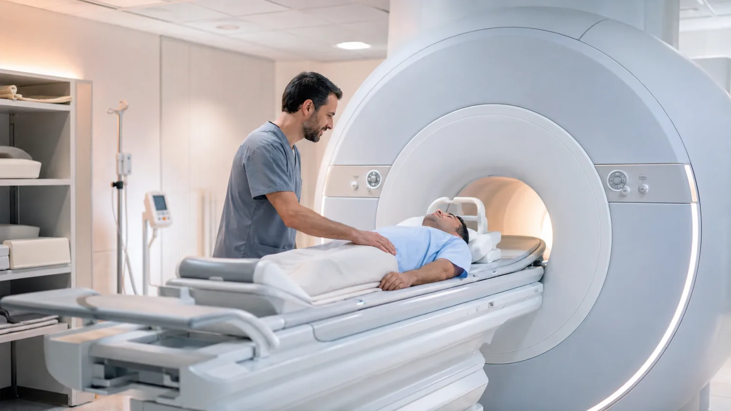
You will usually lie on a motorised table that slides into the scanner. The scanner is shaped like a short tunnel open at both ends. The part of your body being scanned needs to be positioned carefully, and a special receiver coil may be placed around or near the area.
The radiographer will leave the room but can see and hear you. You will usually have a buzzer or call button to press if you need help. You may be given earplugs or headphones because MRI scanners make loud tapping, knocking or buzzing noises.
You need to stay as still as possible so the images are clear. Some scans require you to hold your breath briefly. If you move, parts of the scan may need to be repeated.
The NHS says an MRI scan usually takes 15 to 90 minutes depending on the size of the area being scanned and how many images are taken. NHS: what happens during an MRI scan.
Does MRI hurt?
The scan itself is painless. You do not feel the magnetic field or radio waves. The hardest parts for many people are lying still, staying in position, the noise, and feeling enclosed inside the scanner.
If you have pain when lying flat, tell the radiographer. They may be able to use cushions or supports. If you are having contrast dye, you may feel a brief scratch when the cannula is inserted and sometimes a cool sensation during injection.
Why is MRI so noisy?
MRI scanners make loud knocking, tapping and buzzing sounds because parts of the scanner rapidly switch during image acquisition. This is normal.
You will usually be given ear protection. Some units offer music through headphones, although this depends on the scan and equipment.
What if you are claustrophobic?
Claustrophobia is common. Many people worry about feeling trapped in the scanner. Tell the imaging department before your appointment if you are anxious or have had a difficult MRI before.
Options may include:
- explaining the scan in advance
- keeping communication open throughout
- using mirrors or lighting where available
- music or headphones in some units
- a friend or relative nearby if allowed and safe
- feet-first positioning for some scans
- short breaks between sequences where possible
- mild sedation arranged in advance in selected cases
- open or wider-bore MRI in some centres
Do not take a sedative before MRI unless it has been prescribed and the imaging department knows. If sedation is used, you may need someone to take you home and you should not drive afterwards.
What is MRI contrast dye?
Some MRI scans use a contrast agent, often gadolinium-based, injected into a vein. Contrast can make blood vessels, inflammation, tumours, scars or certain tissues show more clearly.
Contrast may be used for:
- some brain and spine scans
- cancer assessment
- liver or abdominal lesion characterisation
- blood vessel imaging
- infection or inflammation assessment
- some heart scans
- post-surgery assessment
NHS Inform explains that some MRI scans involve contrast injection to make certain tissues and blood vessels show more clearly, and that side effects such as nausea, rash, headache or dizziness are usually mild and short-lived. NHS Inform MRI scan guidance.
Is MRI contrast safe?
MRI contrast is generally safe for most people, but it is not needed for every scan. The team will only use it if it helps answer the clinical question.
Before contrast, you may be asked about:
- kidney disease
- previous reaction to contrast
- pregnancy or breastfeeding
- allergies
- recent blood tests
Serious allergic reactions are rare, but you should tell the team immediately if you feel unwell, itchy, short of breath, dizzy, swollen or develop a rash after contrast.
Can you have MRI if you are pregnant?
MRI may be used during pregnancy if the benefits outweigh any potential risks and the information is needed. It does not use ionising radiation. However, pregnancy should always be discussed with the referring clinician and MRI team, especially if contrast is being considered.
NHS 111 Wales notes that there is no evidence MRI scans are a risk for pregnant women, but whether one is offered depends on why it is needed, and a doctor can discuss whether MRI or contrast is suitable. NHS 111 Wales MRI information.
Can children have MRI?
Yes. Children can have MRI, but the challenge is keeping still. Some children manage well with preparation, explanation and support. Younger children may need sedation or anaesthetic depending on the scan, age and local practice.
Parents or carers should tell the team about implants, previous operations, allergies, anxiety, sensory needs and any possibility of metal fragments or devices.
Who performs and reports an MRI?
The scan is performed by a radiographer. The images are usually reviewed and reported by a radiologist, a doctor specialising in medical imaging. In some specialist areas, reporting may involve other trained professionals or specialist doctors.
The radiologist does not usually discuss results with you on the day. The report is sent to the clinician who requested the scan, such as your GP, consultant or private doctor.
When will you get MRI results?
Timing varies depending on urgency, hospital workload and whether specialist review is needed. Some urgent scans are reported quickly. Routine MRI results may take days to weeks.
Ask before you leave:
- When should I expect the result?
- Who will contact me?
- Should I book a follow-up appointment?
- What should I do if I do not hear anything?
- Are there symptoms that should prompt urgent help before the result arrives?
If you are waiting for a report, see our guide to how to understand scan results.
What might an MRI report say?
MRI reports can be detailed and technical. The report usually describes the body part scanned, the technique, findings, and an impression or conclusion.
Common wording may include:
- No significant abnormality: no important problem seen on the scan.
- Degenerative change: wear-and-tear change, common in joints and spine.
- Disc bulge or protrusion: a spinal disc is bulging, sometimes touching or compressing a nerve.
- Oedema: swelling or fluid signal in tissue or bone marrow.
- Lesion: an area that looks different from surrounding tissue. This does not automatically mean cancer.
- Cyst: a fluid-filled area, often benign depending on features.
- Enhancement: an area takes up contrast dye, which may help characterise it.
- Indeterminate: not clearly benign or serious from MRI alone; follow-up or another test may be needed.
- Correlation advised: the scan finding should be interpreted alongside symptoms, examination or other tests.
Do not interpret a report in isolation. MRI can show incidental findings that are harmless or unrelated to symptoms. Your clinician should explain what the result means for you.
What if the MRI is normal?
A normal MRI can be reassuring, but it does not always mean symptoms are not real. Some conditions are functional, intermittent, microscopic, chemical, inflammatory at a level not visible, or outside the scanned area.
For example, a normal brain MRI does not rule out migraine. A normal spine MRI may not explain all back pain. A normal abdominal MRI may not rule out every digestive condition.
Next steps depend on symptoms. Your doctor may consider blood tests, physiotherapy, nerve tests, medication review, endoscopy, ultrasound, CT, specialist referral or watchful waiting.
Can MRI miss problems?
Yes. MRI is powerful but not perfect. It can miss very small abnormalities, intermittent problems, early disease, movement-affected areas, or problems outside the scanned region. The quality of the scan and the clinical question matter.
This is why red flag symptoms should still be followed up even if an earlier scan was normal. Seek urgent advice for symptoms such as sudden weakness, speech problems, severe new headache, loss of bladder or bowel control with back pain, unexplained weight loss, severe worsening pain, chest pain or severe abdominal pain.
Common finding: degenerative change
Degenerative change means wear-and-tear changes in joints, discs, bones or soft tissues. It is common with age and does not always cause symptoms.
In spine MRI, reports may mention disc dehydration, disc bulges, facet joint arthritis or narrowing. These findings need to be matched with symptoms. Many people without pain have disc or joint changes on MRI.
Common finding: disc bulge
A disc bulge is when a spinal disc extends beyond its usual boundary. It may be mild and incidental, or it may irritate or compress a nerve, causing sciatica, arm pain, numbness, tingling or weakness.
The important question is whether the bulge matches your symptoms and examination. A large-looking bulge may not matter if it is not pressing on a nerve, while a smaller bulge in the wrong place can cause significant symptoms.
Common finding: cyst
Cysts are fluid-filled areas. Many cysts are benign and need no treatment. MRI can help characterise cysts in the liver, kidneys, ovaries, brain, joints and soft tissues.
Some cysts need follow-up if they have complex features, solid parts, thick walls or unusual enhancement. Your clinician will explain whether monitoring or further tests are needed.
Common finding: “incidental” abnormality
An incidental finding is something seen on a scan that was not the reason for the scan. Some incidental findings are harmless. Others need follow-up to make sure they are not important.
Incidental findings can cause anxiety, especially when the report uses technical language. Ask your clinician: Does this finding explain my symptoms? Is it common? Does it need follow-up? What happens next?
Private MRI scans: what to consider
Private MRI scans are widely available. They can be helpful when requested for a clear clinical reason and interpreted in context. However, whole-body or self-referred MRI can sometimes find incidental abnormalities that lead to anxiety, extra tests and unclear benefit.
Before booking privately, ask:
- What question is the MRI trying to answer?
- Is MRI the best test for my symptoms?
- Do I need contrast?
- Who will report the scan?
- Who will explain the result?
- Will my GP receive a copy?
- What happens if something abnormal is found?
- Are follow-up appointments included?
- Will the scan be accepted by NHS or private specialists if I need referral?
If you are using private healthcare, see NHS vs private healthcare in the UK, private GP services and how hospital referrals work.
Whole-body MRI: is it worth it?
Whole-body MRI is sometimes marketed as a proactive health screen. It can detect some serious conditions, but it also has limitations. It may miss important disease, find harmless abnormalities, lead to further tests, or create false reassurance.
For most people, targeted investigation based on symptoms, risk factors, examination and blood tests is more clinically useful than broad scanning without a clear question.
Screening tests are most helpful when there is good evidence they improve outcomes. If you are considering whole-body MRI, discuss the pros and cons with a doctor who is not simply selling the scan.
How to make MRI easier
MRI can be made easier with preparation and communication.
- Read your appointment letter carefully.
- Tell the unit in advance if you have implants, devices or metal fragments.
- Wear comfortable, metal-free clothing.
- Remove jewellery and piercings if possible before attending.
- Tell staff if you are claustrophobic or anxious.
- Use ear protection properly.
- Ask how long the scan will take.
- Use the call button if you need help.
- Focus on slow breathing during the scan.
- Try to keep still so images are clear and sequences do not need repeating.
Questions to ask before or after MRI
Useful questions include:
- Why is MRI being requested?
- What body part is being scanned?
- Will contrast dye be used?
- Do I need kidney blood tests before contrast?
- Do I need to fast?
- Can I take my usual medicines?
- What should I do about implants or devices?
- How long will the scan take?
- When will results be available?
- Who will explain the report?
- What happens if the scan is normal but symptoms continue?
When to seek urgent help instead of waiting for MRI
Some symptoms should not wait for a routine MRI appointment.
Seek urgent medical help if you have:
- sudden weakness, facial drooping, speech difficulty or stroke symptoms
- new loss of bladder or bowel control with back pain
- numbness around the genitals or saddle area
- severe new headache that reaches maximum intensity quickly
- headache with confusion, seizure, weakness or stiff neck
- severe chest pain or breathlessness
- severe abdominal pain or collapse
- fever with severe back pain or neurological symptoms
- rapidly worsening weakness or numbness
- unexplained weight loss with severe worsening symptoms
Call 999 if symptoms suggest stroke, heart attack, severe breathing problems, collapse or another emergency.
Final thoughts
An MRI scan is a detailed imaging test that can show the brain, spine, joints, soft tissues, organs, pelvis, blood vessels and some cancers with excellent detail. It does not use X-rays, but it does use a powerful magnet, so safety screening is essential.
Preparation varies depending on the scan. Many people can eat, drink and take medicines as usual, but some abdominal, pelvic or contrast scans have special instructions. Always follow your appointment letter and tell the MRI team about implants, pacemakers, metal fragments, pregnancy, kidney disease, allergies or claustrophobia.
MRI can provide important answers, but it is not perfect and does not replace clinical judgement. Results should be interpreted alongside symptoms, examination, blood tests and other scans. A normal MRI can be reassuring, while abnormal findings sometimes need careful explanation to avoid unnecessary worry.
For official information, see the NHS guide to MRI scans, NHS Inform’s guide to MRI scans, and UK MRI safety guidance from GOV.UK.
This article is for general information only and should not replace medical advice. Always follow the instructions from your hospital, imaging department or clinician, especially about implants, fasting, medicines, contrast dye and follow-up.
Frequently asked questions
What is an MRI scan?
An MRI scan is a medical imaging test that uses a strong magnetic field and radio waves to create detailed pictures inside the body. It does not use X-rays.
What does MRI show best?
MRI is especially good at showing soft tissues, including the brain, spinal cord, discs, nerves, ligaments, muscles, joints, pelvis, liver, bile ducts, prostate and some cancers.
Does MRI use radiation?
No. MRI does not use ionising radiation. It uses magnets and radio waves.
How long does an MRI take?
Many MRI scans take between 15 minutes and 1 hour, although some take longer depending on the area being scanned and whether contrast is used.
Is an MRI scan painful?
No. MRI itself is painless. You may find it uncomfortable to lie still, and the scanner is noisy. Some people feel anxious or claustrophobic.
What should I wear for MRI?
Wear comfortable clothing without metal if possible. Avoid zips, metal buttons, underwired bras, belts, jewellery and piercings. You may be asked to change into a gown.
Can I eat before an MRI?
Often yes, but it depends on the scan. Some abdominal, pelvic or contrast scans may require fasting. Always follow the instructions in your appointment letter.
Can I take my usual medicines before MRI?
Usually yes, but follow your appointment instructions. If you have diabetes, kidney disease or take medicines that may be affected by fasting or contrast, ask for advice in advance.
What is MRI contrast dye?
MRI contrast is an injection, often gadolinium-based, that helps certain tissues, blood vessels, inflammation or tumours show more clearly. It is not needed for every MRI.
Is MRI contrast dangerous?
MRI contrast is generally safe for most people, but it may not be suitable for everyone. Tell the team if you have kidney disease, allergies, previous contrast reactions, pregnancy or breastfeeding concerns.
Can I have MRI with a pacemaker?
Some modern pacemakers are MRI-conditional, meaning scanning may be possible under strict conditions. Others are not safe. Always tell the MRI team about pacemakers or implanted devices before the scan.
What if I am claustrophobic?
Tell the MRI department before your appointment. They may offer support, communication during the scan, music, positioning adjustments, a wider scanner, or sedation arranged in advance in selected cases.
When will I get MRI results?
Results timing varies. The radiologist reports the scan and sends the report to the clinician who requested it. Ask when to expect results and who will explain them.
Can MRI miss cancer?
Yes. MRI is detailed but not perfect, and it only scans the requested area. A normal MRI does not rule out every cancer or every cause of symptoms.
Is private MRI worth it?
Private MRI can be useful when there is a clear clinical question and proper follow-up. Whole-body or self-referred MRI may find incidental abnormalities, miss some problems or create uncertainty, so it is best discussed with a clinician first.